Medical simulation apparatus and method for controlling 3-dimensional image display in the medical simulation apparatus

a medical simulation and image display technology, applied in the field of medical simulation apparatus and method for controlling 3dimensional image display in the medical simulation apparatus, can solve the problems of occlusion of upper and lower dental arches, inability to check the contact state of teeth, and difficulty in comparing the two-dimensional image information obtained through paper surgery and the three-dimensional image information

Active Publication Date: 2004-12-23
IMAGNOSIS
View PDF14 Cites 21 Cited by
  • Summary
  • Abstract
  • Description
  • Claims
  • Application Information

AI Technical Summary

Benefits of technology

[0013] According to the present invention, the three-dimensional image displayed on the image display means can be changed according to the change in the entity model.
[0022] This method provides the same effects as described in relation to claim 1.

Problems solved by technology

In the surgical orthodontic operation, occlusion of upper and lower dental arches cannot be ignored for positioning of the upper and lower jawbones.
However, it is impossible to check a teeth contact state, because the paper surgery provides only two-dimensional information.
However, it is difficult to reflect and correlate the two-dimensional image information obtained through the paper surgery and the three-dimensional image information obtained through the model surgery with respect to each other.
Therefore, a facial appearance change attributable to the teeth contact state cannot be predicted by the combination of the paper surgery and the model surgery.
However, it is difficult to cut away an image portion of the lower jawbone and displace the cut-away image portion to ensure proper contact between the upper and lower dental arches without any overlap.
This makes it impossible to check the teeth contact state on the display, and to check the facial appearance change attributable to the teeth contact state.
However, it is difficult to reflect and correlate two-dimensional image information obtained through the paper surgery and three-dimensional image information obtained through the model surgery with respect to each other.
However, even if the simulation of the implantation of the dental implant in the dental arch model is performed on the basis of the implantation direction and depth of the dental implant examined on the three-dimensional image, it is impossible to check if the results of the simulation are consistent with the examination on the three-dimensional image.

Method used


Image

  • Medical simulation apparatus and method for controlling 3-dimensional image display in the medical simulation apparatus
  • Medical simulation apparatus and method for controlling 3-dimensional image display in the medical simulation apparatus
  • Medical simulation apparatus and method for controlling 3-dimensional image display in the medical simulation apparatus

Examples


Embodiment Construction

[0030] Embodiments of the present invention will hereinafter be described in detail with reference to the attached drawings.

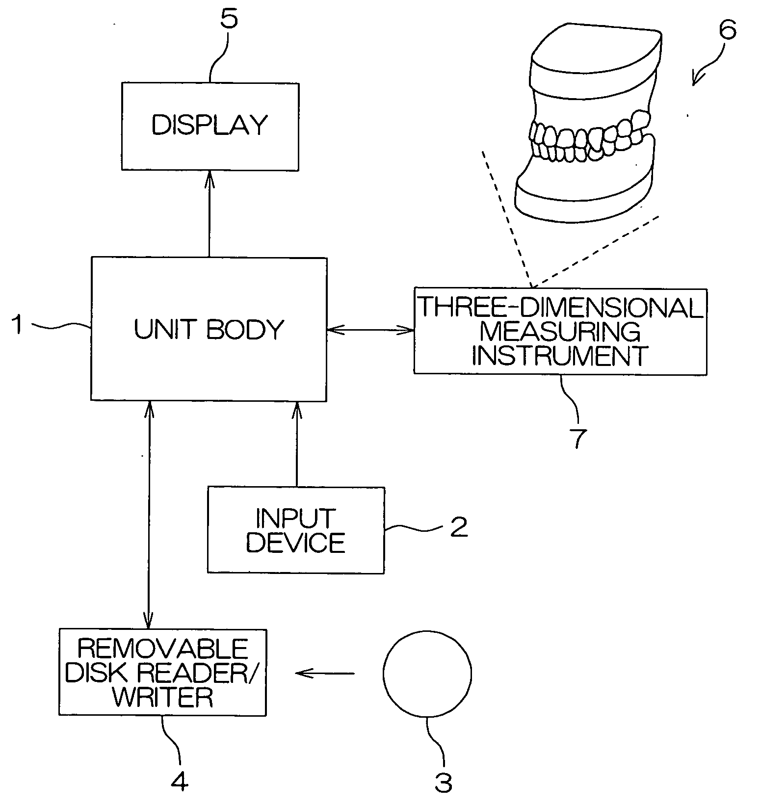
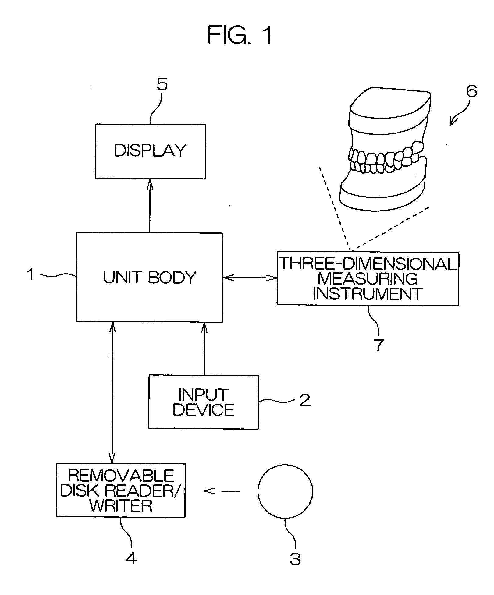
[0031] FIG. 1 is a block diagram illustrating the construction of a medical simulation apparatus according to one embodiment of the present invention. The medical simulation apparatus is adapted to perform a simulation with the use of a three-dimensional image, for example, for a surgical orthodontic operation in dental surgery or plastic surgery, and includes a unit body 1 incorporating a microprocessor and a hard disk therein.

[0032] The unit body 1 is connected to an input device 2 such as a keyboard and a mouse for inputting data and commands, a removable disk reader / writer 4 for writing and reading data with respect to a removable disk 3 such as a DVD (digital video disk), a display 5 for displaying a three-dimensional image, and a three-dimensional measuring instrument 7 for acquiring coordinates of a given point on a surface of an entity model 6 in a coor...


PUM


Abstract

A medical simulation apparatus is provided, which is capable of performing a simulation by correlating a three-dimensional image with an entity model. Coordinates of measurement marks MP1, MP2, MP3 on a surface of a lower dental arch model 62 in a coordinate system defined by three reference marks MK1, MK2, MK3 are acquired before and after displacement of the lower dental arch model 62. The coordinates thus acquired are converted into coordinates in a coordinate system defined by reference marks PK1, PK2, PK3 on the basis of a relationship between the reference marks MK1, MK2, MK3 and the reference marks PK1, PK2, PK3. Then, changes in the coordinates after displacement of the lower dental arch model 62 are determined, and a region preliminarily specified in a three-dimensional image displayed on a display 5 is displaced on the basis of the coordinate changes thus determined.

Description

[0001] The present invention relates to a medical simulation apparatus for performing a simulation of a surgical operation, for example, for confirmation of a surgical technique and prediction of a postoperative state, and to a three-dimensional image display controlling method for the medical simulation apparatus.PRIOR ART[0002] In the medical field, a simulation is often performed with the use of a trace of a radiograph or an entity model (formed of gypsum plaster, a resin or the like). On the basis of the results of the simulation, a surgical technique is confirmed and a postoperative state is predicted. Where a surgical orthodontic operation is performed for treatment of protrusion of an upper jawbone or a lower jawbone (so-called buck teeth or protruding lower lip) or jaw deformity in dental surgery or plastic surgery, for example, a simulation is performed for confirmation of a surgical technique and prediction of a postoperative state.[0003] In the surgical orthodontic operat...

Claims


Application Information

Patent Timeline
Patent Type & Authority Applications(United States)
IPC IPC(8): A61B5/055A61B5/11A61C7/00A61B6/03A61B19/00A61C1/08A61C9/00A61C11/00A61C13/00G01R33/32
CPCA61B5/1116A61B5/1127A61B5/4547A61B19/54A61C1/084A61C7/002A61C9/004A61C11/00A61C13/0004A61C9/0046A61B90/39
Inventor KIM, HAN-JOON
Owner IMAGNOSIS
Why Eureka
Try Eureka