
Blood vessels wall imaging catheter

a blood vessel wall and imaging catheter technology, applied in the field of blood vessel wall imaging catheters, can solve the problems of poor resolution, unmatched by any other imaging modality, and the inability to adapt to atherosclerosis study in large scal

Inactive Publication Date: 2004-06-17
V TARGET TECH
View PDF99 Cites 78 Cited by
  • Summary
  • Abstract
  • Description
  • Claims
  • Application Information

AI Technical Summary

Problems solved by technology

This is unmatched by any other imaging modality.
However, extracorporeal nuclear imaging has not been adapted broadly for study of atherosclerosis due to the radiation and safety problems, poor resolution and lack of specificity, and ordinary gamma probes may not be inserted into the arteries.

Method used


Image

  • Blood vessels wall imaging catheter
  • Blood vessels wall imaging catheter
  • Blood vessels wall imaging catheter

Examples


example 1

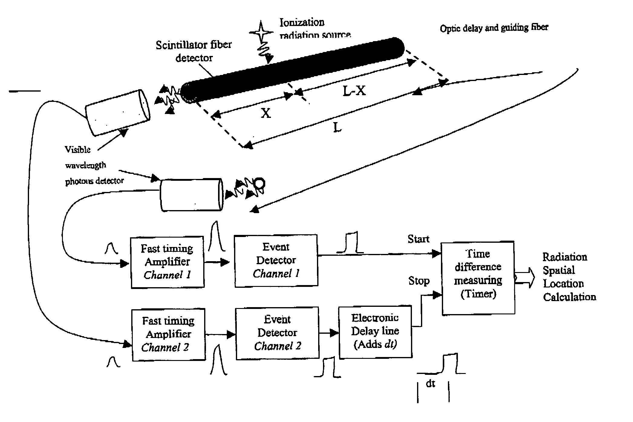
[0052] Gas Filled Detectors with Gas such as CO.sub.2 CH.sub.4

[0053] Ionization chamber

[0054] Proportional chamber

[0055] Geiger chamber

example 2

[0056] Scintillation Detectors

[0057] Organic scintillators crystals and liquids

[0058] C.sub.14H.sub.10, C.sub.14H.sub.12, C.sub.10H.sub.8 etc.

[0059] Plastics

[0060] NE102A, NE104, NE110

[0061] Pilot U

[0062] Inorganic scintillators

[0063] NaI

[0064] CsI

[0065] BGO

[0066] LSO

[0067] YSO

[0068] BaF

[0069] ZnS

[0070] ZnO

[0071] CaWO.sub.4

[0072] CdWO.sub.4

example 3

[0073] Scintillator coupling:

[0074] Photomultiplier Tube (PMT)

[0075] Side-on type

[0076] Head-on type

[0077] Hemispherical type

[0078] Position sensitive

[0079] Microchannel Plate-Photomultiplier (MCP-PMTs)

[0080] Electron multipliers

[0081] Photodiodes (& Photodiodes Arrays)

[0082] Si photodiodes

[0083] Si PIN photodiodes

[0084] Si APD

[0085] GaAs(P) photodiodes

[0086] GaP

[0087] CCD


PUM


Abstract

A system and method are provided for intravascular nuclear imaging, which is furthermore designed to locate the plaque along the artery and to estimate its thickness. Radiolabeled antibodies directed specific antigens in the atherosclerotic plaque or radiolabeled immunoscintigraphy pharmaceutical, which binds apoptotic cells, have been proposed for detection of vulnerable plaque. Other Radiopharmaceuticals such as Gallium Citrate, radio labeled white blood cells etc. call also be used to illuminate inflammatory processes.

Description

[0001] This application derives priority from U.S. Provisional Application No. 60 / 418,712, filed on Oct. 17, 2002, and XXXXX, filed on Nov. 8, 2002.FIELD AND BACKGROUND OF THE INVENTION[0002] The present invention relates to the diagnosis and treatment of cardiac arteries disease, and more particularly, to a system and method for imaging vulnerable plaque in coronary arteries.[0003] Heart attacks seem to occur suddenly and often without warning, yet, the process underlying the event has been going on for many years.[0004] In principle, nuclear imaging is an ideal method to investigate most function processes, mainly perfusion, metabolism and receptor-ligand processes, as it has an exquisite sensitivity being able to identify molecules down to the picomole range. This is unmatched by any other imaging modality. However, extracorporeal nuclear imaging has not been adapted broadly for study of atherosclerosis due to the radiation and safety problems, poor resolution and lack of specifi...

Claims


Application Information

Patent Timeline
IPC IPC(8): A61B6/00
CPCA61B6/00A61B6/4258A61B6/425
Inventor AMRAMI, RONIKIMCHY, YOAV
Owner V TARGET TECH
Learn More