
MR imaging with Dixon-type water/fat separation

a technology of dixon-type water/fat and mr imaging, which is applied in the field of mr imaging with dixon-type water/fat separation, can solve the problems of slow dual-echo acquisition, and poor snr of phase images acquired in the known dixon-type mr imaging methods, so as to achieve high snr, improve noise propagation, and enable dix

Active Publication Date: 2021-01-19
KONINKLJIJKE PHILIPS NV
View PDF10 Cites 0 Cited by
  • Summary
  • Abstract
  • Description
  • Claims
  • Application Information

AI Technical Summary

Benefits of technology

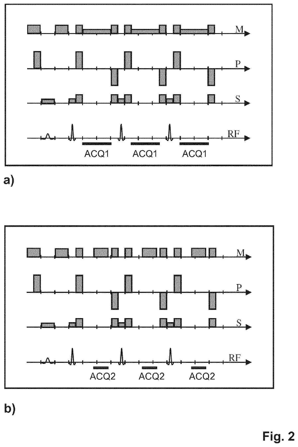
[0019]According to the invention, the number of phase encodings (i.e., the number of k-space lines / profiles) in the acquisition of the in phase signals is smaller than the number of phase encodings in the acquisition of the (partially) out of phase signals. The reduction of the number of phase encodings correspondingly reduces the SNR in an MR image reconstructed from the first (in phase) MR echo signals. This enables tailoring the SNR in the in phase and water (or fat-suppressed) images individually as required, by adapting the SNR in the MR image reconstructed from the first (in phase) MR echo signals relative to the SNR in an MR image reconstructed from the second ((partially) out of phase) MR echo signals. Preferably, the number of phase encodings of the first and second imaging sequences are chosen such that the SNR in the MR image reconstructed from the first (in phase) MR echo signals is essentially equal to the SNR in the MR image reconstructed from the second ((partially) out of phase) MR echo signals.
[0020]In this way, the invention achieves an improved noise propagation in the water / fat separation and, simultaneously, a reduced overall scan time.
[0022]According to a preferred embodiment of the invention, each of the first and second imaging sequences are repeated, partially or entirely, for signal accumulation, wherein the number of repetitions of the first imaging sequence may be different from the number of repetitions of the second imaging sequence. Selectively repeating the first and second imaging sequences respectively can be used in order to further tailor the SNR in the in phase, water, and fat images individually, including improving it in the in phase images and equalizing it in the in phase and water images. Simultaneously, the overall scan time can be kept within acceptable limits.
[0025]As the object is subjected to multiple shots of the first and second imaging sequences, the number of shots of the first imaging sequence may be chosen to be smaller than the number of shots of the second imaging sequence. In this way, a smaller number of phase encodings of the first imaging sequence than the number of phase encodings of the second imaging sequence within the meaning of the invention can be obtained.
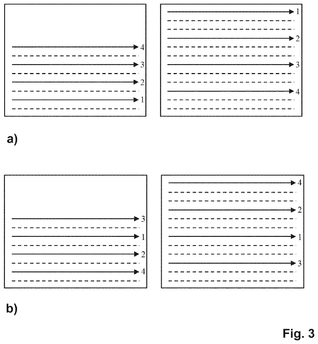
[0026]Preferably, the echo orders of the first and second imaging sequences are chosen such that first and second MR echo signals acquired from a central position in k-space have identical or adjacent echo index numbers, the echo index number indicating the position of each MR echo signal in the train of echo signals generated by each shot of the first or second imaging sequence. By choosing the echo orders in this way, the contrast in the in phase and (partially) out of phase images can be kept as similar as possible.
[0028]In a preferred embodiment of the invention, the phase encoding scheme of the first imaging sequence is chosen such that k-space is incompletely sampled using a partial Fourier technique. By applying a partial Fourier technique to the acquisition of the first MR echo signals a corresponding reduction of the number of phase encodings of the first imaging sequence in comparison to the number of phase encodings of the second imaging sequence can be obtained. The partial Fourier technique exploits the symmetry of the MR signal data in k-space to reduce the number of phase encodings (and correspondingly the required acquisition time) by sampling only an asymmetric part of k-space. The symmetry in k-space is a basic property of Fourier transformation and is called Hermitian symmetry. Thus, from the known symmetry properties for the MR signal data a full MR image can be reconstructed from the MR signal data from only one half of k-space. The conditions under which Hermitian symmetry holds and the corrections that must be applied when the assumption is not strictly obeyed must be considered. A prerequisite for partial Fourier imaging is that phase shifts are slowly varying across the object. In this regard, the invention takes advantage from the typically very smooth spatial variation of the phase in the in phase TSE image data. However, to avoid problems due to slowly varying phase shifts, a little more than one half of k-space should be covered by the first imaging sequence. Preferably, both (positive and negative) sides of k-space (in the phase encoding direction) are acquired in a low spatial frequency range while at higher spatial frequencies the MR signal data are acquired only on one side. The fully sampled low frequency portion can be used to characterize (and correct for) any slowly varying phase shifts.

Problems solved by technology

However, constraints on the echo times may actually render dual-echo acquisitions slower than triple-echo acquisitions.
The in phase and out of phase images acquired in the known Dixon-type MR imaging methods suffer from a poor SNR as compared to the water and fat images obtained with these methods.
Moreover, the in phase images acquired in the known Dixon-type MR imaging methods suffer from a poor SNR as compared to in phase images acquired by means of separate, tailored, non-Dixon-type MR imaging methods.
A comparable SNR would often only be achievable in prohibitively long scan times.
However, a drawback of this known approach is that the variation of the receiving bandwidth leads to an unequal SNR in the two sub-acquisitions and thus to an unfavorable noise propagation in the water / fat separation.

Method used


Image

  • MR imaging with Dixon-type water/fat separation
  • MR imaging with Dixon-type water/fat separation
  • MR imaging with Dixon-type water/fat separation

Examples


Embodiment Construction

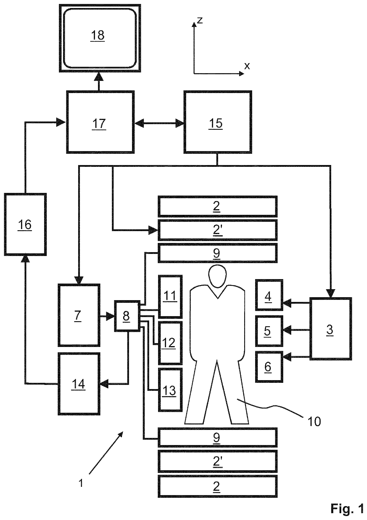
[0035]With reference to FIG. 1, a MR device 1 is shown. The device comprises superconducting or resistive main magnet coils 2 such that a substantially uniform, temporally constant main magnetic field B0 is created along a z-axis through an examination volume. The device further comprises a set of (1st, 2nd, and—where applicable—3rd order) shimming coils 2′, wherein the current flow through the individual shimming coils of the set 2′ is controllable for the purpose of minimizing B0 deviations within the examination volume.

[0036]A magnetic resonance generation and manipulation system applies a series of RF pulses and switched magnetic field gradients to invert or excite nuclear magnetic spins, induce magnetic resonance, refocus magnetic resonance, manipulate magnetic resonance, spatially and otherwise encode the magnetic resonance, saturate spins, and the like to perform MR imaging.

[0037]More specifically, a gradient pulse amplifier 3 applies current pulses to selected ones of whole-...


PUM


Abstract

The invention relates to a method of Dixon-type MR imaging. It is an object of the invention to provide a method that enables Dixon water / fat separation with high SNR and with improved noise propagation in the water / fat separation. The method comprises the steps of: —subjecting an object (10) to a first imaging sequence, which generates a number of differently phase encoded first MR echo signals at a first echo time, such that contributions from MR signals emanating from water protons and MR signals emanating from fat protons to the first MR echo signals are essentially in phase, —acquiring the first MR echo signals using a first signal receiving bandwidth, —subjecting the object (10) to a second imaging sequence which generates a number of differently phase encoded second MR echo signals at a second echo time, such that contributions from MR signals emanating from water protons and MR signals emanating from fat protons to the second MR echo signals are at least partially out of phase, —acquiring the second MR echo signals using a second signal receiving bandwidth which is larger than the first receiving bandwidth, wherein the number of phase encodings of the first imaging sequence is smaller than the number of phase encodings of the second imaging sequence, and —reconstructing a MR image from the first and second MR echo signals, whereby signal contributions from water protons and fat protons are separated. Moreover the invention relates to a MR device and to a computer program to be run on a MR device.

Description

CROSS REFERENCE TO RELATED APPLICATIONS[0001]This application is a U.S. national phase application of International Application No. PCT / EP2017 / 080286 filed on Nov. 24, 2017, which claims the benefit of EP Application Serial No. 16200449.3 filed on Nov. 24, 2016 and is incorporated herein by reference.FIELD OF THE INVENTION[0002]The invention relates to the field of magnetic resonance (MR) imaging. It concerns a method of MR imaging of a portion of a body placed in the examination volume of a MR device. The invention also relates to a MR device and to a computer program to be run on a MR device.BACKGROUND OF THE INVENTION[0003]Image-forming MR methods which utilize the interaction between magnetic fields and nuclear spins in order to form two-dimensional or three-dimensional images are widely used nowadays, notably in the field of medical diagnostics, because for the imaging of soft tissue they are superior to other imaging methods in many respects, do not require ionizing radiation ...

Claims


Application Information

Patent Timeline
Patent Type & Authority Patents(United States)
IPC IPC(8): G01R33/48G01R33/54G01R33/561
CPCG01R33/4828G01R33/543G01R33/5611G01R33/5617G01R33/561G01R33/5615
Inventor EGGERS, HOLGER
Owner KONINKLJIJKE PHILIPS NV
Learn More