
Method and apparatus for adjustment of a table position in a medical data acquisition scanner

a technology of medical data acquisition and table position, which is applied in the direction of instruments, applications, and measurements using nmr, can solve the problems of incompatibility of scanning protocols for different body regions, one scanning protocol cannot be used to acquire image data in different body regions, etc., and achieves the effect of partial flexibility

Active Publication Date: 2020-08-18
SIEMENS HEALTHCARE GMBH
View PDF19 Cites 0 Cited by
  • Summary
  • Abstract
  • Description
  • Claims
  • Application Information

AI Technical Summary

Benefits of technology

The invention allows for a scanning protocol to be applied to different body regions and the position of the table to be set independently of the scanning protocol. This means that the table can be positioned based on the anatomical information of the body region being examined or to minimize table movements. This flexibility improves image quality and reduces the number of table movements, resulting in improved efficiency and accuracy of medical data acquisition. Additionally, the use of magnetic resonance medical data acquisition scanners, which require the user to stipulate the body region being examined under time pressure, can also benefit from this flexibility in selecting a scanning protocol.

Problems solved by technology

One disadvantage with this, however, is that for different body regions, for example, for different body regions of a human body or different organs, a specialized scanning protocol is often required since, for example, the contrast ratios in the different regions may vary to such an extent that one scanning protocol cannot be used to acquire image data in different body regions.
In other words, scanning protocols are incompatible for different body regions.

Method used


Image

  • Method and apparatus for adjustment of a table position in a medical data acquisition scanner
  • Method and apparatus for adjustment of a table position in a medical data acquisition scanner
  • Method and apparatus for adjustment of a table position in a medical data acquisition scanner

Examples


Embodiment Construction

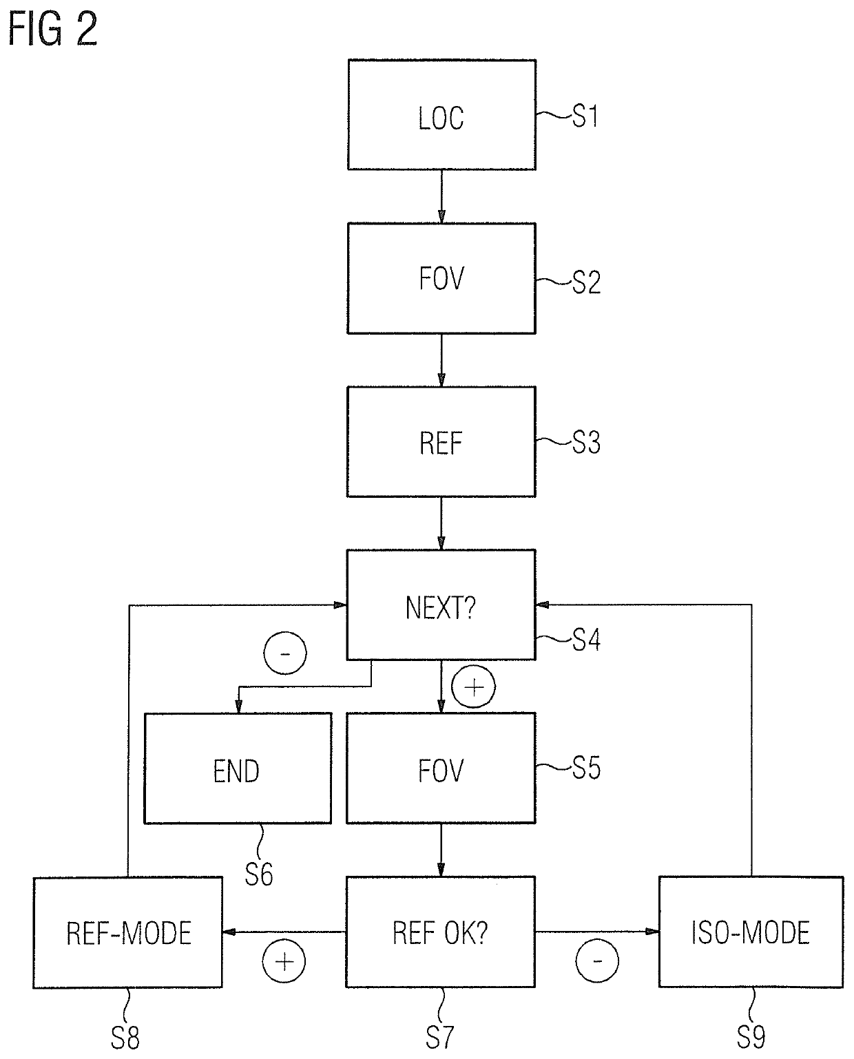
[0034]The exemplary embodiment described hereinafter is a preferred embodiment of the invention. In the exemplary embodiment, however, the components of the embodiment that are described each represent individual features of the invention that are to be considered independently of one another, which in each case further develop the invention independently of one another and are thus also to be seen as a component of the invention either individually or in a different combination from that shown. Furthermore, the embodiment described can also be complemented by further features of the invention that have already been described.

[0035]In the figures, elements with an identical function are each denoted by the same reference signs.

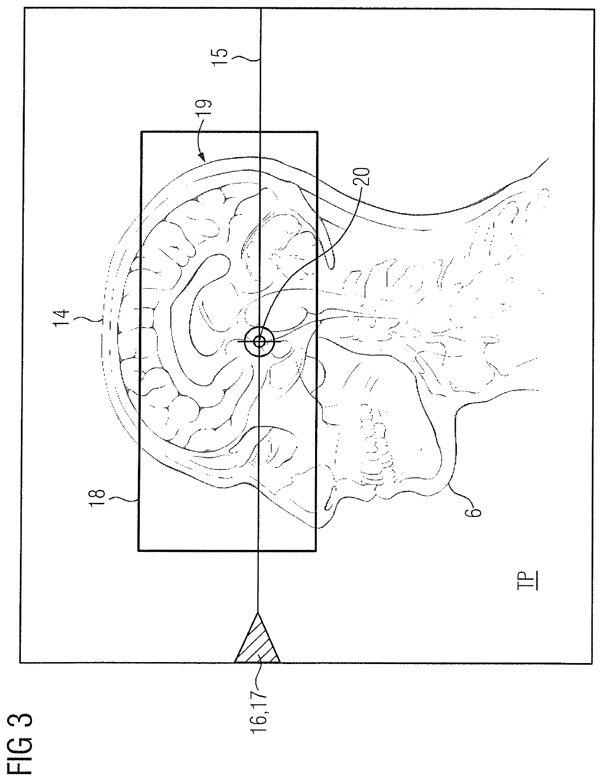
[0036]FIG. 1 shows an apparatus that can be installed, for example, in a hospital or a medical center or a radiology center. The apparatus 1 comprises a medical data acquisition scanner 2, a table 3, a control device 4, and optionally an operating device 5. Th...


PUM


Abstract

In a method and apparatus for adjusting a table position in a medical data acquisition scanner, image data relating to a body region are generated by operation of the medical data acquisition scanner by running a scanning protocol. The scanning protocol is intended to be used independently of the body region that is mapped. An iso-mode, in which a relative position of the body region relative to the isocenter of the medical data acquisition scanner can be set via the table position in accordance with an optimization criterion, and on a ref-mode, in which the table position is set at a predetermined reference position, are provided. A mode of operation is selected from the iso-mode and the ref-mode as dependent on anatomical information relating to the body region and / or a relative position of the body region relative to the isocenter that ensues in the reference position, and before the running of and / or during the running of the selected scanning protocol, the table position is set according to the selected mode of operation.

Description

BACKGROUND OF THE INVENTIONField of the Invention[0001]The invention concerns a method for operating a control device, in order to adjust a table position of a table relative to a medical data acquisition scanner. A body arranged on the table is intended to be examined by the medical data acquisition scanner. The invention also includes an apparatus having a medical data acquisition scanner and the control device that is operated according to the invention.Description of the Prior Art[0002]As used herein, an examination means the acquisition of image data for a body region of the body by operation of the medical data acquisition scanner. Such an examination or session begins with a body, for example a (human or animal) patient or a material sample of a material (for example of a metal or a rock), being placed on the table and the table then being moved into a table position or a table position being set, at which the body region that is to be examined has a favorable position relati...

Claims


Application Information

Patent Timeline
Patent Type & Authority Patents(United States)
IPC IPC(8): G06F3/0484A61B5/00G01R33/30G01R33/54A61B5/055G06F3/0482G01R33/28
CPCG06F3/04842A61B5/7435G06F3/04847G06F3/0482A61B5/0555G01R33/28A61B5/704G01R33/546A61B6/0407A61D3/00A61B5/055
Inventor KEIL, MIRIAMKRELLMANN, CHRISTOFSCHMITT, PETER
Owner SIEMENS HEALTHCARE GMBH
Learn More