
Methods for the isolation of extracellular vesicles and other bioparticles from urine and other biofluids

a technology of bioparticles and vesicles, applied in the field of cell biology, can solve the problems of prohibitive co-purification of pcr inhibitors and complicating downstream nucleic acid analysis, and achieve the effect of not co-purifying prohibitive amounts of pcr inhibitors

Active Publication Date: 2020-06-02
YMIR GENOMICS
View PDF5 Cites 1 Cited by
  • Summary
  • Abstract
  • Description
  • Claims
  • Application Information

AI Technical Summary

Benefits of technology

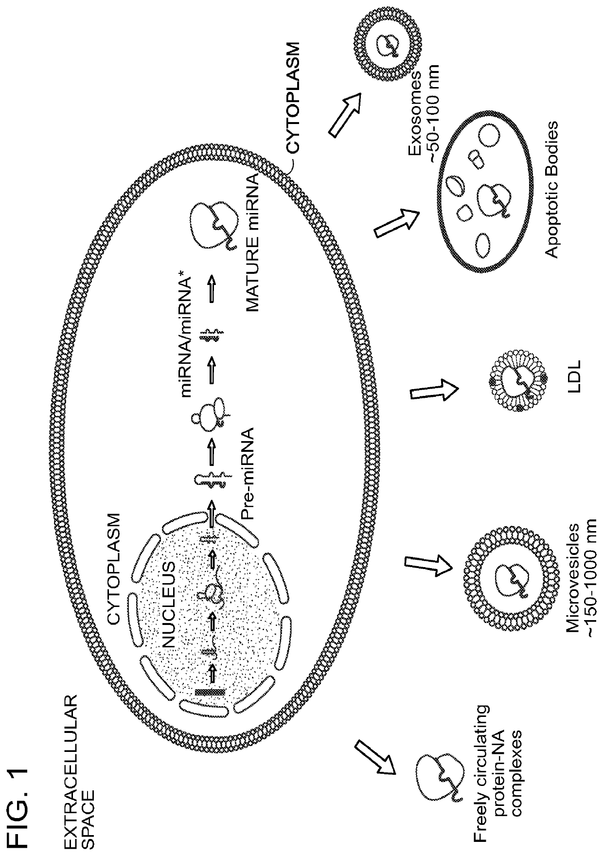
[0017]The current invention is based, at least in part, upon discovery of a means for isolating bioparticles from liquid sample (e.g., biofluid) using several approaches, including a crystal-promoting and / or precipitation method and an apparent matrix-binding method that is optionally suitable for columns (without wishing to be bound by theory, the matrix-binding method appears to exploit pore sizes of certain materials to effect enrichment, such as the pore sizes found in porous beads, e.g., siliceous beads such as diatomaceous earth and perlite). In certain aspects, the invention provides methods for the rapid and inexpensive isolation of bioparticles: specifically, membrane-bound vesicles, cell-free protein-nucleic acid complexes, cell-free mRNA, and / or cell-free DNA can be isolated from almost any fluid. These methods use common laboratory equipment and reagents. They do not require high-speed centrifugation, such as ultracentrifugation. They do not require expensive membranes, antibodies, antibody fragments, beads, or sophisticated columns. Such methods produce a higher yield of bioparticles and known bioparticle markers than many other methods. The methods do not co-purify prohibitive amounts of PCR inhibitors that would complicate downstream nucleic acid analysis. In some embodiments, the methods allow for isolation of intact microvesicles, enabling mechanistic, delivery, vaccine-related, immunostimulation-related and therapeutic downstream studies.
[0019]In certain embodiments, the invention provides methods for isolating released bioparticles from whole urine samples, where those methods comprise i) treating whole urine samples with the reducing agent TCEP (tris(2-carboxyethyl)phosphine, optional; TCEP protects against the loss of microvesicles in the subsequent low speed spin), ii) spinning the urine samples in a low speed spin (typically 1000×g for typically 5 minutes) to remove cellular contamination and debris (contained in the pellet), iii) applying the crystal and / or precipitation inducing reagent Monosodium Urate to the supernatant of the previous spin, iv) incubating the mixture, typically on ice or 4 degrees and typically for 15 minutes, v) centrifuging the mixture to form a pellet and a supernatant, most advantageously, in a low speed centrifugation, vi) removing the supernatant after the spin and, vii) recovering the pellet by resuspending in a resuspension solution.
[0027]The invention is superior to ultracentrifugation methods because i) it does not require an expensive ultracentrifuge, ii) it is significantly faster, iii) it does not lose as many microvesicles in the first centrifugation step, and iv) as judged by some markers for urine microvesicles and extracellular miRNA, has a higher yield, especially in more dilute urine samples (see FIGS. 4 and 5).
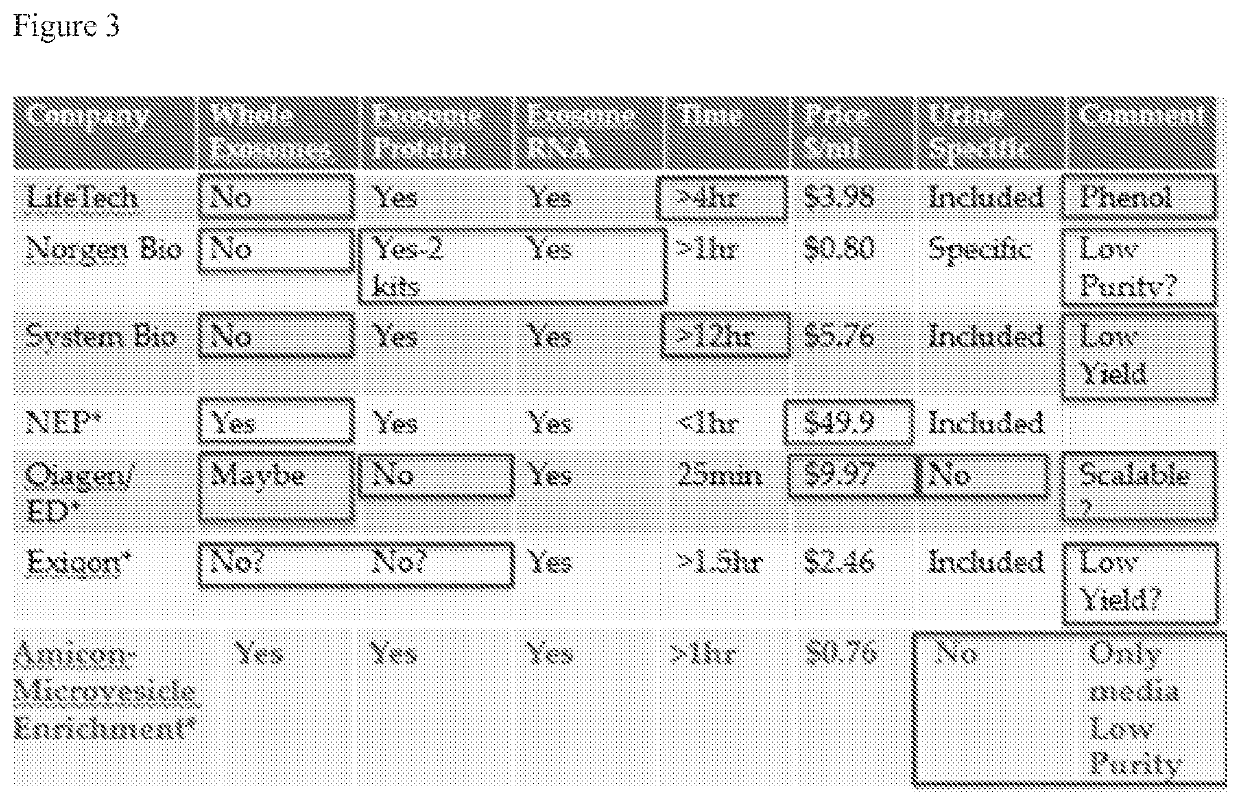
[0028]The invention is also superior to existing commercial and academic precipitation methods in that i) it does not lose as many microvesicles in the first centrifugation step (see FIG. 20), ii) the incubation time is significantly shorter, iii) the crystal / precipitation-inducing agent or the porous beads are significantly less expensive than other precipitation-inducing reagents, and iv) as judged by some markers for urine microvesicles, has a higher yield, especially in more dilute samples (see FIGS. 4 and 5).
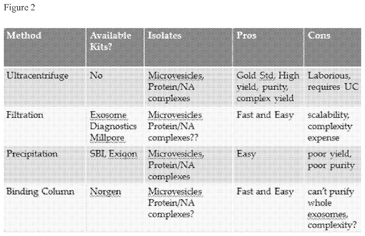
[0029]Certain embodiments of the invention are superior to existing precipitation, column and filtration methods, in that i) they do not lose as many microvesicles in the first centrifugation step, ii) do not require expensive column housing, column packing, or filters, iii) can be significantly faster than, iv) can be easily scaled up to large volumes of biofluid and v) as judged by some markers for urine microvesicles and extra-cellular miRNA, have a higher yield.
[0054]An additional aspect of the invention provides a method for reducing the microvesicle content of a liquid sample from a subject or cell culture, the method involving: a) obtaining a liquid sample from a subject or cell culture; b) contacting the liquid sample with a crystal / precipitation-inducing agent under conditions suitable to allow for crystal formation and / or precipitation, thereby creating an admixture; c) incubating the admixture for a period of time sufficient to allow for crystal formation and / or precipitation; d) separating the admixture to obtain a particle fraction and a liquid fraction and isolating the liquid fraction, thereby reducing the microvesicle content of a liquid sample from a subject or cell culture.

Problems solved by technology

The methods do not co-purify prohibitive amounts of PCR inhibitors that would complicate downstream nucleic acid analysis.

Method used


Image

  • Methods for the isolation of extracellular vesicles and other bioparticles from urine and other biofluids
  • Methods for the isolation of extracellular vesicles and other bioparticles from urine and other biofluids
  • Methods for the isolation of extracellular vesicles and other bioparticles from urine and other biofluids

Examples


example 1

A Newly Discovered Na Urate Protocol Isolated Microvesicles from Urine Quickly and Effectively

[0176]To exemplify certain methods of the invention, 3 mls whole urine samples from two different donors (one sample was naturally concentrated and one sample was naturally dilute) were treated with 16 mM TCEP reducing agent as part of a Whole Urine Prespin Treatment Solution, which simultaneously reduced the pH to <6 and was believed to have reduced the matrix-forming properties of the abundant endogenous urine protein, THP. The mixture was immediately centrifuged at 1,000×g for 5 minutes to remove cells and debris. The supernatant was gently removed and then 40 microliters per ml of sample of 131 mM Monosodium urate (in 1 N NaOH) was added to create a mixture. This mixture was incubated for 15 minutes on ice and then centrifuged for 5 minutes at 1,000×g in a desktop microcentrifuge. After centrifugation, the supernatant was gently removed and the pellet was resuspended in a small volume o...

example 2

The Na Urate Process More Completely Depleted Urine of Bioparticles than the Ultracentrifuge Method

[0187]The fact that the instant Na Urate method isolated significantly more of several protein and microRNA markers for bioparticles, and also of particles as judged by NTA and TEM (see Example 1 above), strongly suggested that the instant method could isolate the same bioparticles which the heretofore gold standard method of ultracentrifugation could. This was important, as there was also value in depleting biofluids such as urine, blood serum / plasma, and tissue culture serum of bioparticles. To determine if the instant method more completely depleted urine of bioparticles than the ultracentrifuge method, the instant method and the ultracentrifuge method were applied to 1.5 mls of urine from the same sample. Subsequently, the respective final supernatants for each method represented bioparticle-depleted urine. These depleted urine samples were then applied to the alternate method (i.e...

example 3

The Na Urate Process Isolated Bioparticles / Microvesicles from Non-Urine Biofluids

[0188]To determine if the instant method could isolate bioparticles from liquid other than urine, bioparticles were initially isolated from 1.5 ml of urine using ultracentrifuge. These bioparticles were then added to pure water, and the instant method was applied. This was considered to be an ideal test for the hypothesis that the instant method could isolate bioparticles from other fluids, because water contains no salt, has a neutral pH, and also has no other constituents of urine. As FIG. 15 shows, the instant method was capable of isolating a small amount of TSG101 and a significant amount of CD9 exosomal markers even from water (lane 6). Although little Aqua-2 was recovered, this was likely due to the small amount of Aqua-2 isolated by ultracentrifuge in the first place (lane 3), which meant that little Aqua-2 was introduced into the water at the outset. This demonstrated the ability of the instant...


PUM

PropertyMeasurementUnit
pore sizeaaaaaaaaaa
pore sizeaaaaaaaaaa
pore sizeaaaaaaaaaa

Abstract

Compositions and methods for the isolation of protein-nucleic acid complexes and microvesicles (collectively referred to as “bioparticles”) released by mammalian cells into body fluids or cell culture media are provided. Isolated bioparticles of the invention contain biological molecules that are useful as diagnostic / prognostic biomarkers or for identification of therapeutic targets (e.g., disease or disorder-associated miRNAs). The isolation of biological molecules as described herein results in purification and concentration of the molecules. Methods for producing bio fluids that are free of detectable bioparticles, that are largely depleted of bioparticles, or that possess a reduced concentration of bioparticles compared to a bio fluid starting material (collectively termed “bioparticle-depleted”) are also provided. Isolation of bioparticle-depleted biofluid is useful, e.g., in experimental systems where it is desirable to use a biofluid that does not contain endogenous bioparticles, or has been substantially depleted of endogenous bioparticles, from the source material.

Description

CROSS-REFERENCE TO RELATED APPLICATIONS[0001]This application is the U.S. national stage pursuant to 35 U.S.C. § 371, of United States International Application Ser. No. PCT / US2015 / 043768, filed Aug. 5, 2015 and published in English on Feb. 11, 2016 as publication WO2016 / 022654 A1, which is related to U.S. Provisional Patent Application Ser. No. 62 / 033,643, entitled “Methods for the Isolation of Cell-Free Protein-Nucleic Acid Complexes and Microvesicle Bioparticles from Liquids”, which was filed Aug. 5, 2014, and to U.S. Provisional Patent Application Ser. No. 62 / 033,644, entitled “Methods for the Isolation of Cell-Free Protein-Nucleic Acid Complexes, Biomarkers and Microvesicle Bioparticles from Urine”, which was filed Aug. 5, 2014. The entire contents of these patent applications are hereby incorporated by reference herein.FIELD OF THE INVENTION[0002]The invention relates to the field of cell biology, and in particular, to the study of circulating, cell-free, membrane-bound struct...

Claims


Application Information

Patent Timeline
Patent Type & Authority Patents(United States)
IPC IPC(8): C12N15/10B01D21/26B01D9/00G01N1/40C07K1/30C07K14/705
CPCC07K1/30B01D9/0054B01D21/26C07K1/306C07K14/705C12N15/1003G01N1/4055C07K1/303B01D2009/0086B01D21/262
Inventor PENDERGRAST, PATRICK SHANNONPENDERGRAST, ROBERT SCOTTPENDERGRAST, JOHN STEPHENMARKOWSKA, ANNA IRMINA
Owner YMIR GENOMICS
Learn More