
Methods for measuring and reporting vascularity in a tissue sample

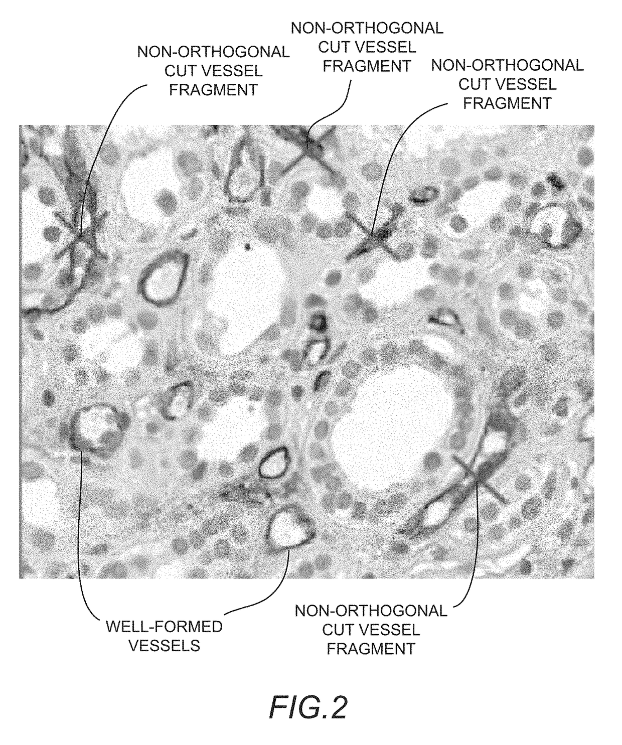
a tissue sample and vascularity technology, applied in the field of tissue sample vascularity measurement and reporting, can solve the problems of one vessel, difficult for pathologists (and especially the computer) to determine which vessel profiles, and inability to adequately number the vessels with image analysis

Active Publication Date: 2019-01-29
FLAGSHIP BIOSCI
View PDF8 Cites 0 Cited by
  • Summary
  • Abstract
  • Description
  • Claims
  • Application Information

AI Technical Summary

Benefits of technology

The patent describes methods for identifying cells near a vessel, based on their oxygenation level. This approach is not dependent on the number of vessels in the area, but instead measures the distance of the cells to the nearest vessel. The percentage of target tissue that is within or outside of this distance is determined, and reported. The methods can be used for identifying cells in various tissues and diseases, such as multiple myeloma. Overall, the patent offers a novel approach for analyzing the microenvironment of cells in vivo.

Problems solved by technology

This is made difficult in that it must either be studied: (i) non-invasively, using radiologic or other non-destructive imaging modalities; (ii) evaluated with histopathology using tissue sections; or (iii) with lab assays with two-dimensional and three-dimensional cell cultures.
There are both theoretical and experimental problems with this endpoint.
When one considers microvessel density from a stereological viewpoint, recognizing that a two-dimensional tissue section is only one sample from the three dimensional tumor, a number of theoretical problems present themselves.
Experimentally, the difficulty with vessel densities is the ability to adequately number vessels with image analysis.
In tumors with limited vascularity consisting of only small microcapillaries, it may be possibly, but as vascularity increases, it becomes difficult for the pathologist (and especially the computer) to determine which vessel profiles should be part of only one vessel.
This assumes that large, non-oxygenating vessels are removed (an important software quality control technique recommended by the applicant) and also still suffers from the challenge of adequately assigning endothelial cells to vessel counts.
The existing microvessel density techniques reviewed above each suffers from the difficult of computationally assigning cells to vessels.
This difficulty has resulted in limited usage of these techniques in clinical samples.
Another disadvantage is that these techniques report the number or area of vessels, when biologically what may be more appropriate is the percentage of a given cell type that is actually near a vessel.

Method used


Image

  • Methods for measuring and reporting vascularity in a tissue sample
  • Methods for measuring and reporting vascularity in a tissue sample

Examples


Embodiment Construction

[0032]In the following description, for purposes of explanation and not limitation, details and descriptions are set forth in order to provide a thorough understanding of the embodiments of the invention. However, it will be apparent to those skilled in the art that the present invention may be practiced in other embodiments, including certain variations or alternative combinations that depart from these details and descriptions.

[0033]The methods disclosed herein are enhanced with certain aspects of the methods for feature analysis on consecutive tissue sections, as are described in commonly owned U.S. Pat. No. 8,787,651 issued Jul. 22, 2014; the contents of which are hereby incorporated by reference.

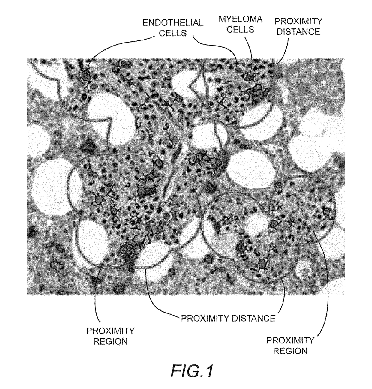
[0034]For purposes of this disclosure, the term “proximity analysis” refers to the determination of an amount of a first cellular entity that is within a given distance of a second distinct cellular entity;

[0035]“microvessel proximity analysis” refers to proximity analysis where one of ...


PUM


Abstract

The disclosure concerns a method for measuring and reporting vascularity in a biological tissue sample. The method generally includes: within a digital image of a tissue section, (i) identifying endothelial cells, lymphatic cells, or a combination thereof; (ii) mapping one or more proximity regions, each of the proximity regions defining an area between detected vessels and a first distance outwardly therefrom; and (iii) calculating one or more of: a vessel proximity score or a hypoxia score, wherein the vessel proximity score relates a composition of objects within the proximity regions, and wherein the hypoxia score relates a composition of tissue within or outside of the proximity regions, respectively.

Description

CROSS-REFERENCE TO RELATED APPLICATIONS[0001]This application is a continuation of U.S. Ser. No. 14 / 588,319, filed Dec. 31, 2014, titled “METHODS FOR MEASURING AND REPORTING VASCULARITY IN A TISSUE SAMPLE”;[0002]which claims benefit of priority with U.S. Provisional Ser. No. 62 / 030,518, filed Jul. 29, 2014;[0003]the entire contents of each of which are hereby incorporated by reference.BACKGROUND OF THE INVENTIONField of the Invention[0004]This invention relates to methods for measuring and reporting vascularity in a tissue sample; and more particularly, to methods for evaluation of angiogenesis and hypoxia using digital image analysis platforms.Related Art[0005]The ability to evaluate the vascular structure of tissue is important in many therapeutic areas, both in areas that seek to increase the growth of blood vessels (pro-angiogenic) and areas that seek to decrease or shrink blood vessels (anti-angiogenic). It is important to be able to evaluate the architecture of the blood vascu...

Claims


Application Information

Patent Timeline
Patent Type & Authority Patents(United States)
IPC IPC(8): G06T7/00G06K9/00G01N33/50G01N33/569
CPCG01N33/56966G01N33/5044G01N33/5064G01N33/56972G06K9/00127G06T7/0012G06T2207/30101G01N2800/7014G01N2800/7038G06T2207/10024G06T2207/10056G06T2207/30024G06V20/69
Inventor POTTS, STEVEN J.
Owner FLAGSHIP BIOSCI
Learn More