
Non-invasive system and method for monitoring lusitropic myocardial function in relation to inotropic myocardial function

a myocardial function and non-invasive technology, applied in the field of non-invasive systems and methods for monitoring cardiac parameters, can solve the problems of patient well along the way down the slippery slope of decompensation and death, no solution that allows for non-invasive measurement of lusitropy, and no one has solved the problem, so as to achieve the effect of inexpensively, safely, reliably diagnosing and monitoring hfpef, and reducing the risk of cardiac death

Active Publication Date: 2018-10-02
THE COOPER HEALTH SYST
View PDF8 Cites 1 Cited by
  • Summary
  • Abstract
  • Description
  • Claims
  • Application Information

AI Technical Summary

Benefits of technology

[0012]A. The sepsis studies show that in sepsis, lusitropy, in relation to inotropy, declines in a sentinel fashion, well before these other metrics. This new invention gives clinicians more time to intervene.
[0013]B. Similarly, in the context of myocardial ischemia, an ST-segment depression is a late sign of an imbalance between oxygen supply and demand, in favor of demand. It is well known that regional wall motion abnormalities (“RWMAs”) on a two dimensional echocardiographic image of the left ventricle in cross section precede ST-segment depression by several minutes. It is likely that a decline in lusitropic function precedes RWMAs in the context of myocardial ischemia. That would give clinicians a longer window of opportunity in which to restore oxygen supply and demand balance, and allow them to prevent myocardial ischemia and infarction in the first place.
[0014]C. Noninvasive measurement of the relation between lusitropy and inotropy can serve to diagnose and monitor heart failure and HFpEF, also known as diastolic heart failure. At present, diastolic heart failure is diagnosed when a patient has signs and symptoms of heart failure, but has a normal ejection fraction on two dimensional echocardiogram. Alternatively, an elevated BNP level in the presence of a normal ejection fraction or stress echocardiogram is necessary to make the diagnosis. The present invention represents a cheaper, more reliable way to make the diagnosis and quantify the natural history and treatment of HFpEF. It uses real time physiologic metrics rather than imaging or laboratory values of blood biomarker concentrations.
[0020]By allowing lusitropic function to be quantified non-invasively, the present invention can serve to inexpensively, safely, and reliably diagnose and monitor HFpEF, as well as the efficacy, or inefficacy of therapeutic medications and procedural interventions for cardiac problems across the spectrum. It is also key to quantifying chronic conditions characterized by poor lusitropic function, such as diastolic heart failure, or HFpEF, and its several causes, such as hypertension, diabetes, aortic stenosis, constrictive pericarditis, and restrictive cardiomyopathy, including amyloidosis, sarcoidosis and fibrosis. By allowing lusitropic function to be quantified non-invasively, the present invention can serve to inexpensively, safely, and reliably diagnose and monitor HFpEF, as well as the efficacy, or inefficacy of therapeutic medications and procedural interventions. The technology can serve in one embodiment as a bedside monitor in hospital, or, combined with smartphone and Internet connectivity, the invention can also serve in another embodiment as a new window into myocardial function in the emerging world of telemedicine, helping to deliver better, faster, cheaper, safer care, in a way that radically increases access to care for large communities of people who are not now being well served by the existing health care system. This telemedical measurement, characterization, and cloud storage of the patient's ‘Physiome’ data, owned by the patient, is well described in Dr. Eric Topol's recent book, ‘The Patient Will See You Now’ (Basic Books, 2015).

Problems solved by technology

Researchers have worked to develop non-invasive methods to measure cardiac output and indices of preload and afterload; however, to date there is no solution that allows for non-invasive measurement of lusitropy, except as it relates to acute myocardial ischemia, where lusitropy rapidly and dramatically decreases, and only then, as shown by the depression or elevation of the ST segment of an EKG with ischemia.
Nor has anyone solved the problem of determining in a non-invasive way the beat-by-beat relationship between inotropy and lusitropy as an index of myocardial well-being or pathology.
The overriding issue is that by the time these other metrics, such as blood pressure, cardiac output, or stroke volume, start to diminish acutely, the patient is already well along his way down the slippery slope of decompensation and death.
The procedure done there is hazardous, with risk for fatal arrhythmias, infection, and hemorrhage.
It uses expensive disposables, and generates a great deal of infectious waste, whose disposal is expensive.
It is certainly not at all well suited to clinical monitoring, let alone telemedicine.

Method used


Image

  • Non-invasive system and method for monitoring lusitropic myocardial function in relation to inotropic myocardial function
  • Non-invasive system and method for monitoring lusitropic myocardial function in relation to inotropic myocardial function
  • Non-invasive system and method for monitoring lusitropic myocardial function in relation to inotropic myocardial function

Examples


Embodiment Construction

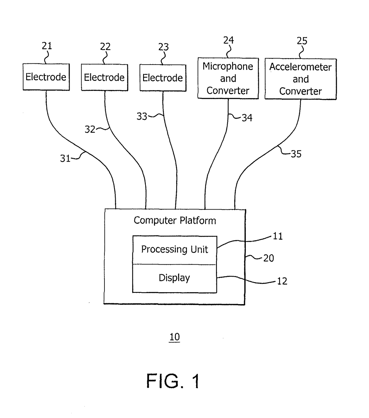
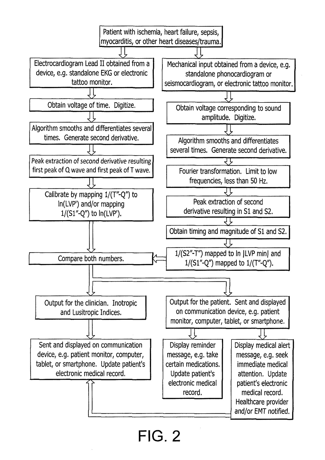
[0023]One object of this invention is to provide an inexpensive, safe, continuous, non-invasive, metric based on lusitropic function, and lusitropic function relative to inotropic function on a beat-to-beat basis, that is determined in a non-invasive manner, that allows for continuous monitoring in the emerging smartphone-connected telemedicine space. Another objective is to improve access to care by removing traditional obstacles to clinical assessment and diagnosis by measuring a physiologic cardiodynamic quantity that is not now being measured on a non-invasive basis, whose decompensation results in at least three categories of disease: myocardial ischemia, sepsis, and heart failure, whose costs are huge and whose consequences are devastating. The system of the present invention is comprised of at least one electrocardiographic lead containing an electrode, preferably lead II, but it should be noted that the preferred embodiment employs greater accuracy by using three leads havin...


PUM


Abstract

A system and method for non-invasively monitoring the hemodynamic state of a patient by determining on a beat-by-beat basis the ratio of lusitropic function to inotropic function as an index of myocardial well-being or pathology for use by clinicians in the hospital or by the patient at home. In one embodiment of the system a smartphone running an application program that is connected through the internet to the cloud processes electronic signals, first, from an electrocardiogram device monitoring electrical cardiac activity, and second, from a seismocardiogram device monitoring mechanical cardiac activity in order to determine such ratio as an instantaneous measurement of the hemodynamic state of the patient, including such states as sepsis, myocardial ischemia, and heart failure.

Description

CROSS REFERENCE TO RELATED APPLICATION[0001]This application claims the benefit of U.S. Provisional Application No. 62 / 238,330 filed Oct. 7, 2015.FIELD OF THE INVENTION[0002]The instant invention relates to a non-invasive system and method to monitor cardiac parameters, specifically lusitropic myocardial function in relation to inotropic myocardial function on a beat by beat basis in order to assess myocardial well-being or pathology on a real time basis.BACKGROUND OF THE INVENTION1. The Problem to be Solved[0003]Lusitropic function is the ability of the left ventricle to relax during diastolic filling, following maximal left ventricular tension, and ejection of the stroke volume with each heartbeat during systole. Inotropic function is the ability of the myocardium to develop tension and pressure in the left ventricle during systole. Quantifying lusitropic function, and lusitropic function in relation to inotropic function, is the key to quantifying acute conditions, such as myocar...

Claims


Application Information

Patent Timeline
Patent Type & Authority Patents(United States)
IPC IPC(8): A61B5/0452A61B5/04A61B5/00A61B5/044A61B5/11A61B7/04
CPCA61B5/0452A61B5/002A61B5/0022A61B5/044A61B5/1107A61B7/04A61B5/04017A61B2562/0219A61B5/7257G16H40/67Y02A90/10A61B5/316A61B5/339A61B5/349
Inventor HIRSH, ROBERT ALAN
Owner THE COOPER HEALTH SYST
Learn More