
Synthetic vaginal cuff model and method of simulating vaginal cuff closure

a vaginal cuff and model technology, applied in the field of surgical training medical devices, can solve the problems of insufficient training, foregoing disclosure, and the conventional art as a whole, failing to provide a safe and effective manner of practicing

Active Publication Date: 2018-09-25
UNIV OF SOUTH FLORIDA
View PDF13 Cites 0 Cited by
  • Summary
  • Abstract
  • Description
  • Claims
  • Application Information

AI Technical Summary

Benefits of technology

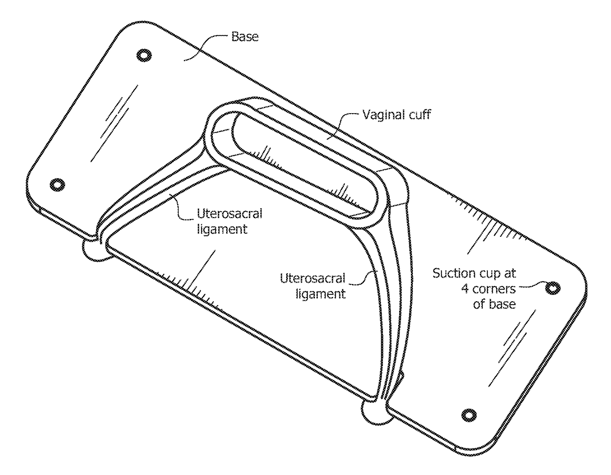
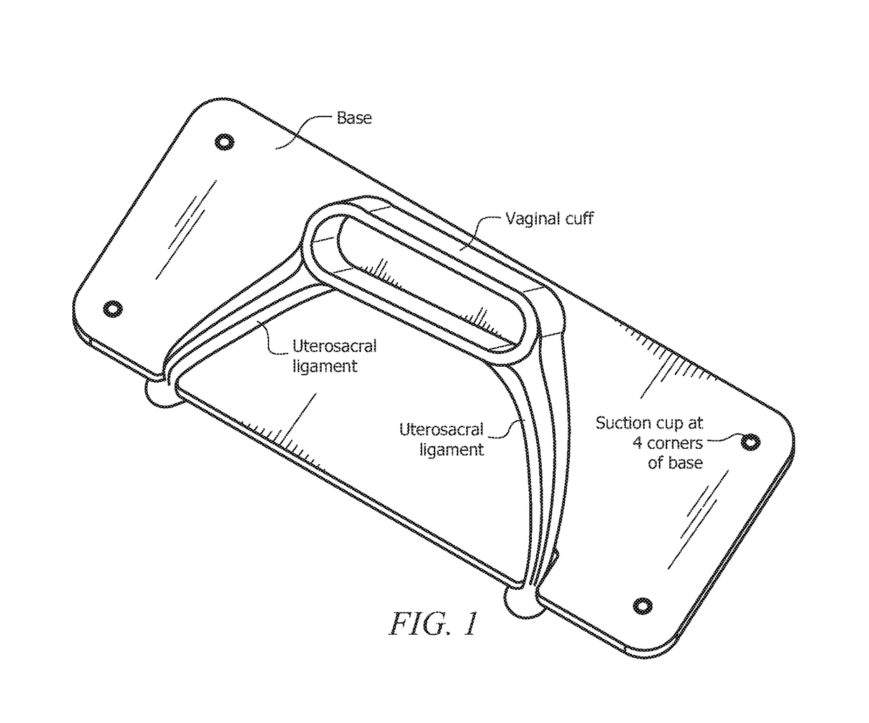
The invention is a new and useful vaginal cuff model and method of training for a cuff closure procedure. The model includes a rounded body resembling a vaginal cuff of an animal, with a hollow interior and at least one open end, and an appendage resembling a uterosacral ligament extending from the rounded body. The model is made of a polymeric material with an embedded layer of mesh disposed along its outer surface. The mesh can be embedded about 1-2 mm below the outer surface of the rounded body and / or appendage. The model can also include a resin or liquid silicone binder. The method of manufacturing the model involves providing a mold for the rounded body and a mold for the appendage, and embedding layers of mesh into both polymeric materials. The model can be used for training purposes by placing it on a stand with a base and an angled component, and attaching a suture pad to it for use in a simulated vaginal cuff closure surgical procedure. The invention addresses a need for a more effective and efficient training tool for this surgical procedure.

Problems solved by technology

However, to perform complex post-hysterectomy surgical techniques, such as reattaching and / or cardinal ligaments to support the vaginal cuff and prevent vaginal vault prolapse, sufficient training is required, as many complications can arise.
The foregoing disclosures, along with the conventional art as a whole, fail to provide a safe and effective manner of practicing post-hysterectomy surgical techniques, particularly when attempting to reattach the uterosacral and / or cardinal ligaments to support the vaginal cuff.
Furthermore, where a definition or use of a term in a reference, which is incorporated by reference herein, is inconsistent or contrary to the definition of that term provided herein, the definition of that term provided herein applies and the definition of that term in the reference does not apply.

Method used


Image

  • Synthetic vaginal cuff model and method of simulating vaginal cuff closure
  • Synthetic vaginal cuff model and method of simulating vaginal cuff closure
  • Synthetic vaginal cuff model and method of simulating vaginal cuff closure

Examples


example

Cuff Closure Simulation

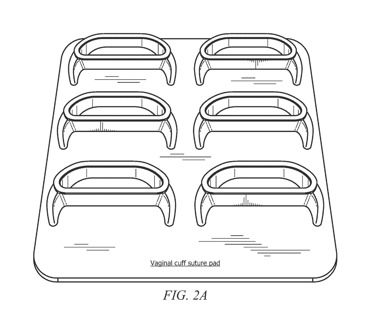
[0044]FIGS. 4A-4B depict the vaginal cuff suture pad of FIGS. 2A-2B as disposed on a hinged, simulation stand that has multiple stable, angled positions to maintain the suture pad at an angle relative the x axis. To simulate closure of a vaginal cuff, a six-cuff model is positioned on the suture pad stand. The stand is angled to the desired / preferred level; optionally, there can be three (3) distinct angled positions (as can be seen in the figures) or it can be stable at any angle. Different angles can provide for different types of training and additional challenges for the surgeon.

[0045]Optionally, the stand can be placed within a laparoscopic (e.g., SIMSEI Laparoscopic Trainer) to simulate the abdominal cavity. Alternatively, the stand can be placed upon a table or other flat surface for simulating the procedure. Once in the desired location, the suction cups (not shown in these figures but indicated in FIG. 1), can be pressed down to hold the stand and sut...


PUM


Abstract

A vaginal cuff surgical model and methods of manufacture and use thereof. The model includes a body (resembling a vaginal cuff) and appendages (resembling uterosacral ligaments) extending from the body. The model (body and appendages) is formed of silicone, rubber, a polymer, or other suitable material with one or more layers of mesh embedded within the polymeric material. The embedded mesh permits suture between the components of the model. The surgical model permits surgical practice of this important component of a total hysterectomy procedure via suturing across the model and to the appendages.

Description

CROSS-REFERENCE TO RELATED APPLICATIONS[0001]This nonprovisional application is a continuation of and claims priority to U.S. Provisional Patent Application No. 62 / 186,961, entitled “Synthetic Vaginal Cuff Model”, filed Jun. 30, 2015 by the same inventors, the entirety of which is incorporated herein by reference.BACKGROUND OF THE INVENTION[0002]1. Field of the Invention[0003]This invention relates, generally, to medical devices for surgical training. More specifically, it relates to methods and apparatuses for modelling vaginal cuffs and simulating surgical procedures pertaining to vaginal cuffs.[0004]2. Brief Description of the Prior Art[0005]Methods and apparatuses are known in the art for supporting vaginal cuffs, particularly after a hysterectomy and / or for the treatment or prevention of prolapse. For example, U.S. Pat. No. 8,636,643 to Kovac et al teaches a device for use in supporting posterior vaginal tissue in a patient without a uterus. The device includes implants that co...

Claims


Application Information

Patent Timeline
Patent Type & Authority Patents(United States)
IPC IPC(8): G09B23/28G09B23/30
CPCG09B23/30G09B23/281
Inventor SIMOES, MARIO ALVESHART, STUART RICHARDCARAMBOT, OMARARMSTRONG, MARK EVANS
Owner UNIV OF SOUTH FLORIDA
Learn More