
Immunohistochemistry division bore dying paste, methods employed and purpose of the same

A technology of immunohistochemistry and tissue sectioning, which is applied in the preparation of test samples, material inspection products, biological testing, etc., to save immunohistochemical reagents and improve work efficiency

Inactive Publication Date: 2007-05-16
郑智勇
View PDF0 Cites 5 Cited by
  • Summary
  • Abstract
  • Description
  • Claims
  • Application Information

AI Technical Summary

Problems solved by technology

However, it is not possible to use the immunohistochemical method of drawing circles to perform multiple immunohistochemical stains on a small area of ​​cell smear; multiple immunohistochemical staining

Method used


Image

  • Immunohistochemistry division bore dying paste, methods employed and purpose of the same
  • Immunohistochemistry division bore dying paste, methods employed and purpose of the same

Examples


Embodiment Construction

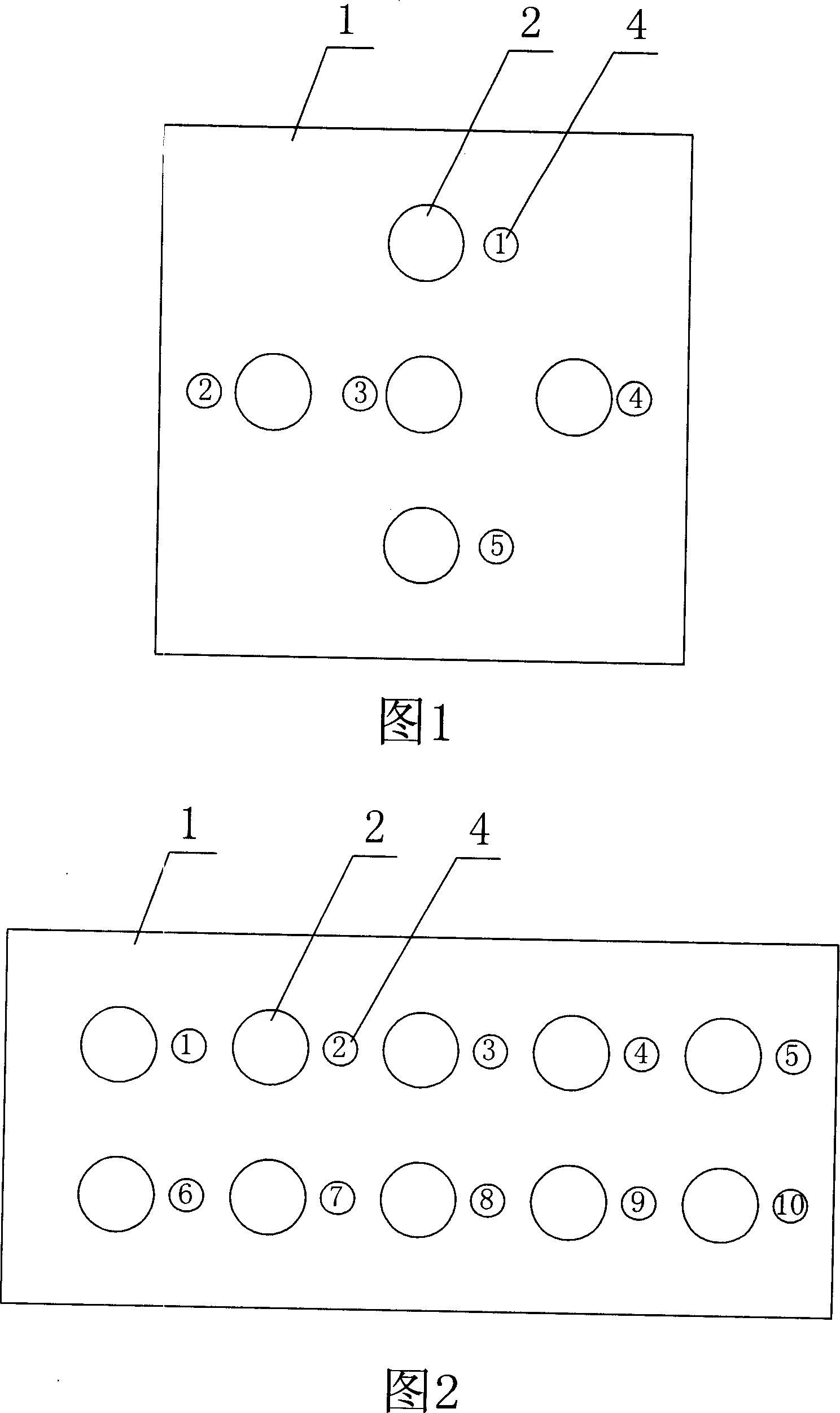
[0018] As shown in Figure 1 or Figure 2, it is a well-made immunohistochemical staining patch, which includes a film or sheet 1 with an area smaller than a slide glass; more than one penetrating small hole 2 is distributed on the film or sheet, so The hole described above is preferably a circular hole, with a certain distance between the hole and the edge of the film or sheet; a self-adhesive is coated on the bottom surface of the above-mentioned film or sheet; and on the above-mentioned The serial number 4 of the hole is printed around the hole of the film or sheet. The film or sheet is preferably made of plastic.

[0019] In order to facilitate the preservation of the prepared immunohistochemical staining patch, the bottom surface of the above-mentioned film or sheet is pasted on the anti-adhesive paper 3, as shown in FIG. 3 .

[0020] The steps of using the above-mentioned immunohistochemical staining patch for multiple immunohistochemical staining are as follows: first pe...


PUM


Abstract

This invention relates to immune group hole dye slice and its usage method, which comprises the following usage steps: sticking tightly more than one transparent holes film slices into load slice coated with organic coupon and dripping immune agent into each hole form immune group dying and the agent volume is limited to the hole edge; ripping off film slices or dipping the load slice into organic liquid to make film peeled off for sealing.

Description

technical field [0001] The invention relates to an immunohistochemical split-hole staining patch, its use method and application. Background technique [0002] In the past, when performing immunohistochemical staining on cell smears or tissue sections on glass slides, in order to control the staining range, it was necessary to first draw a circle on the cell smear or tissue sections with an immunohistochemical circle pen, and then draw a circle around the circle. Immunohistochemical staining was performed. However, it is not possible to use the immunohistochemical method of drawing circles to perform multiple immunohistochemical stains on a small area of ​​cell smear; Multiple immunohistochemical stains were performed. In pathological work, it is often encountered that there is only a small area of ​​cell smear, or there is only one tissue section, but multiple immunohistochemical stains need to be performed on it. Contents of the invention [0003] In order to solve th...

Claims


Application Information

Patent Timeline
IPC IPC(8): G01N1/30G01N33/53
Inventor 郑智勇
Owner 郑智勇
Learn More