
A left ventricle inner and outer membrane automatic segmentation method, system and equipment

A technology for automatic segmentation and left ventricle, applied in image analysis, image data processing, instruments, etc., can solve the problems of accuracy and time performance to be improved, and achieve the effect of good stability, universal applicability and high accuracy

Active Publication Date: 2021-07-23
SOUTH CENTRAL UNIVERSITY FOR NATIONALITIES
View PDF14 Cites 2 Cited by
  • Summary
  • Abstract
  • Description
  • Claims
  • Application Information

AI Technical Summary

Problems solved by technology

Therefore, for many segmentation algorithms, their accuracy and time performance still need to be improved.

Method used


Image

  • A left ventricle inner and outer membrane automatic segmentation method, system and equipment
  • A left ventricle inner and outer membrane automatic segmentation method, system and equipment
  • A left ventricle inner and outer membrane automatic segmentation method, system and equipment

Examples


Embodiment 1

[0130] Adopt the method described in the present invention to extract the left ventricular endocardium, then use the official algorithm evaluation code provided by the MICCAI2009 database to evaluate the segmentation effect. The evaluation index used is the above-mentioned Good Detect percentage, referred to as Good and Overlap in the following tables. Data format: mean (Mean) and standard deviation (SD). Table 3-1 is a comparison between the algorithm in this paper and Lu's algorithm. The research object is 15 cases in the data source, and the compared data is the good detection rate of the endocardium obtained by the segmentation of the two algorithms. The effectiveness and accuracy of the algorithm are reflected by a good detection rate. It can be seen from Table 3-1: the good detection rate obtained by using Lu's algorithm is: 72.45±18.86%; the good detection rate obtained by using this algorithm is: 82.29±16.47%. High robustness.

[0131] Table 3-1 Comparison of detect...

Embodiment 2

[0138] The watershed algorithm based on marker control was used to extract the left ventricle epicardium contour from 15 cases of MICCAI2009 data source, and the segmentation results were analyzed and compared. Figure 16 It is an example of the segmentation result of the epicardial contour in the diastolic image of a case of myocardial enlargement (SC-HYP-06) using the watershed algorithm based on marker control. Each diastolic MRI shows two images, the binary image of the region of interest on the left and the segmentation result on the right. The solid lines are the gold standard of the adventitia, our automatically segmented contours, and the inner and outer markers, respectively. For cases of myocardial enlargement, compared with other images, external markers cannot be extracted directly based on empirical values, so this type of image is processed separately, and the range of external markers is appropriately expanded to avoid excessive indentation of the extracted epic...


PUM


Abstract

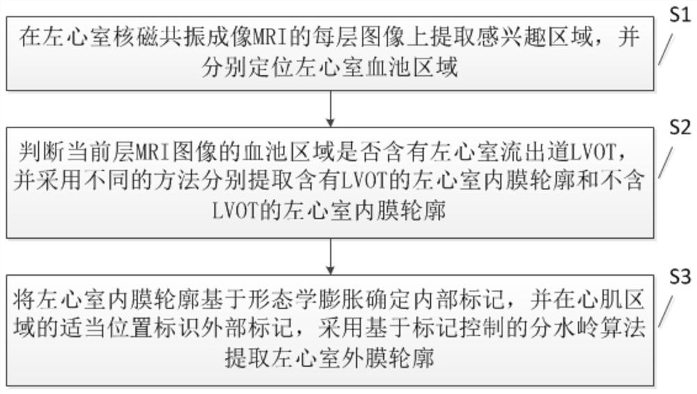
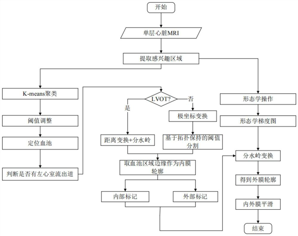
The invention provides a left ventricle inner and outer membrane automatic segmentation method, system and equipment, and belongs to the technical field of nuclear magnetic resonance. The method comprises the following steps: extracting a region of interest on each layer of image of left ventricle magnetic resonance imaging (MRI), and respectively positioning a left ventricle blood pool region; judging whether the blood pool area of the MRI image of the current layer contains a left ventricular outflow tract LVOT or not, and respectively extracting a left ventricular intima contour containing the LVOT and a left ventricular intima contour not containing the LVOT by adopting different methods; and determining an internal mark of the left ventricular intima contour based on morphological expansion, marking an external mark at a proper position of the myocardial area, and extracting the left ventricular adventitia contour by adopting a watershed algorithm based on mark control. By using the method provided by the invention, the accuracy of segmenting the left ventricular inner and outer membranes is high, and the stability and the universality are good.

Description

technical field [0001] The present invention relates to the field of nuclear magnetic resonance technology, in particular to a method, system and equipment for automatic segmentation of left ventricular endocardium and adventitia. Background technique [0002] Early diagnosis and treatment of cardiovascular diseases can reduce mortality to a large extent, which is crucial to prolonging human life. With the continuous development and wide application of modern medical imaging technology, high-definition, high-resolution, multi-information, and low-noise cardiac images provide clinicians with better data sources. At present, in the diagnosis of cardiovascular diseases, medical imaging examinations have been widely used, mainly including: cardiovascular angiography, electrocardiogram (ECG), echocardiography (cardiac color Doppler ultrasound), cardiac magnetic resonance image (Cardiac MRI) and computer Tomography (Computed Tomography, CT), etc. Among these techniques, angiogra...

Claims


Application Information

Patent Timeline
Patent Type & Authority Applications(China)
IPC IPC(8): G06T7/00G06T7/11G06T7/187G06T5/00G06T5/30
CPCG06T7/0012G06T7/11G06T7/187G06T5/30G06T2207/10088G06T2207/20152G06T2207/30048G06T5/70
Inventor 王新年
Owner SOUTH CENTRAL UNIVERSITY FOR NATIONALITIES
Learn More