
Method for collecting fundus images and fundus camera

A fundus image and camera technology, applied in the fields of collecting fundus images, methods and fundus cameras, can solve the problems of poor imaging quality of handheld/portable fundus cameras, unsatisfactory effects of small pupils, etc., to reduce the time spent on shooting, and the shooting process is simple and convenient , good user experience

Pending Publication Date: 2020-12-01
南京览视医疗科技有限公司
View PDF6 Cites 0 Cited by
  • Summary
  • Abstract
  • Description
  • Claims
  • Application Information

AI Technical Summary

Problems solved by technology

[0004] Aiming at the above-mentioned technical problems in the prior art, the embodiment of the present application proposes a method for collecting fundus images and a fundus camera to solve the unsatisfactory effect of capturing small pupils by desktop fundus cameras in the prior art, and the imaging problems of hand-held / portable fundus cameras. Poor quality and other issues

Method used


Image

  • Method for collecting fundus images and fundus camera
  • Method for collecting fundus images and fundus camera
  • Method for collecting fundus images and fundus camera

Examples


Embodiment Construction

[0031] In the following detailed description, numerous specific details of the application are set forth by way of example in order to provide a thorough understanding of the relevant disclosure. It will be apparent, however, to one skilled in the art that the application may be practiced without these details. It should be understood that the terms "system", "device", "unit" and / or "module" used in this application are used as a means to distinguish between different components, elements, parts or assemblies at different levels in a sequential arrangement. method. However, these terms may be replaced by other expressions if the same purpose can be achieved by other expressions.

[0032] It will be understood that when a device, unit or module is referred to as being "on," "connected to" or "coupled to" another device, unit or module, it can be directly on the other device, unit or module. connected or coupled to or communicate with other devices, units or modules, or interv...


PUM


Abstract

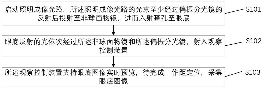
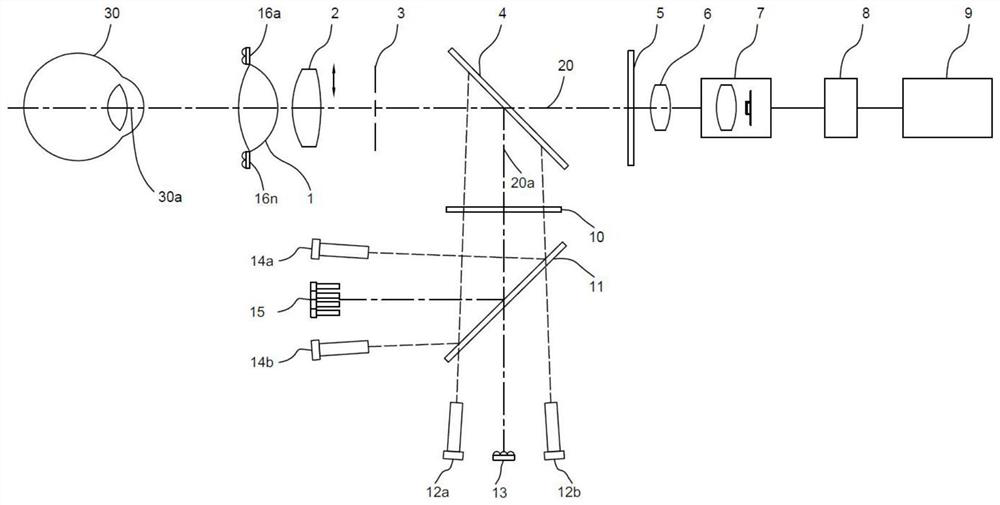
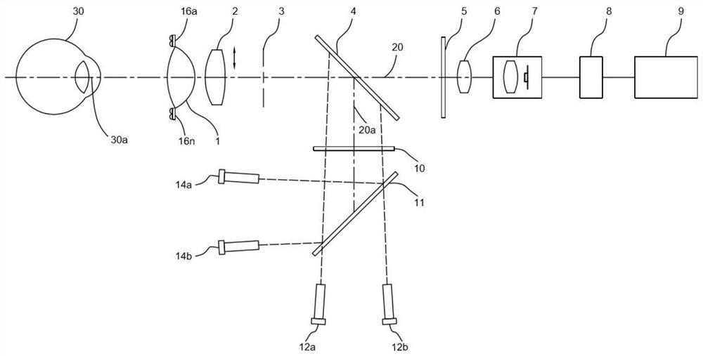
The embodiment of the invention discloses a method for collecting fundus images and a fundus camera, and relates to the technical field of medical ophthalmic optical instruments. The method specifically comprises the following steps: an illumination imaging light path is started, and a light beam of the illumination imaging light path is at least reflected by a polarizing beam splitter, then is projected to an aspheric objective lens and then enters pupils to the fundus; light reflected by the fundus sequentially passes through the aspheric objective lens and the polarizing beam splitter and enters an observation control device; and the observation control device supports real-time preview of fundus images, and the fundus images are collected after the work distance positioning is completed. By the adoption of the method, self-adaption to the pupil size is achieved, the observed preview images are consistent with the shot fundus images, adjustment can be conducted at any time accordingto the preview effect, shooting time is shortened while shooting quality is guaranteed, the whole shooting process is simple and convenient, imaging quality is high, and user experience is good.

Description

technical field [0001] The present application relates to the technical field of medical ophthalmic optical instruments, in particular to a method for collecting fundus images and a fundus camera. Background technique [0002] The fundus camera is a kind of medical ophthalmology examination equipment, which is used to observe and obtain the fundus image of the human eye. Using the fundus camera, the doctor can observe whether the retina, optic disc and blood vessel distribution of the fundus are abnormal and lesions. It can also be used for diabetic retinopathy and glaucoma. Early screening and diagnosis of various diseases such as macular degeneration. [0003] Common desktop fundus cameras on the market can support shooting with a pupil size above 3.3mm at most. Since the pupil size cannot be judged before shooting, it needs to be constantly changed according to the pupil size. For example, when the pupil is smaller than 4.0mm, it needs to be changed from The 4.0mm large ...

Claims


Application Information

Patent Timeline
Patent Type & Authority Applications(China)
IPC IPC(8): A61B3/12A61B3/14G03B15/02
CPCA61B3/12A61B3/14A61B3/0008G03B15/02
Inventor 沈小厚赖明曲志敏许冉冉刘向阳
Owner 南京览视医疗科技有限公司
Learn More