


Quick Research
Generate reliable direction feasibility study reports for your R&D in just a few steps.

Technical Q&A
Discover and master advanced knowledge NOW. Basics, ideas, possibilities, all at once.

Find Solutions
As an expert in R&D theories, this can generate solutions to your technical problems instantly.

Evaluate Feasibility
Analyze your overall solution with one click, know your potential R&D risks in advance.

Monitor Landscape
Get weekly tech updates, stay abreast of the latest tech innovations and key insights.
Three-dimensional image detection system applied to operating room and imaging method using same
A 3D image and detection system technology, applied in the field of medical imaging, can solve problems such as inability to operate in the operating room, huge body size, and difficulty in disinfection, and achieve the effects of easy reconstruction of 3D images, flexible operation, and reduced radiation range
- Summary
- Abstract
- Description
- Claims
- Application Information
AI Technical Summary
Problems solved by technology
Method used
Image


Examples
Embodiment 1
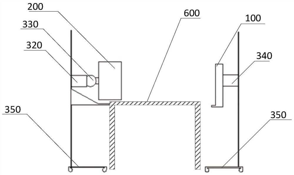
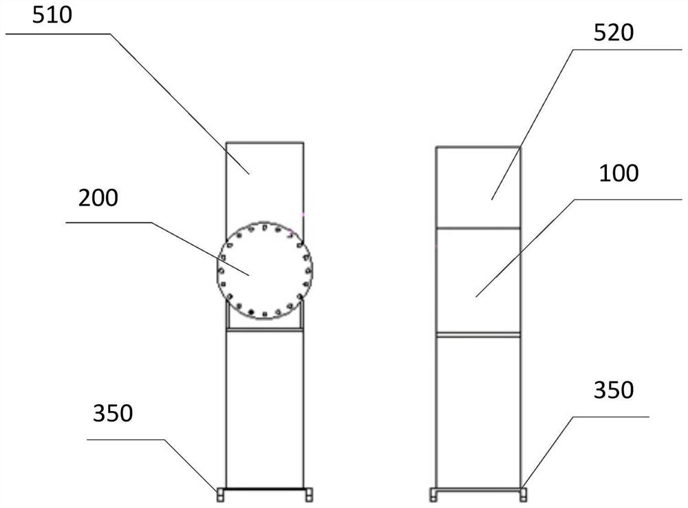
[0049] Reference attached Figure 1-5 As shown, this embodiment provides a three-dimensional image detection system applied to an operating room, which is applied to an operating table 600; including an X-ray receiver 100, an X-ray source 200 and a supporting device. It also includes an imaging scanning device 300 and an X-ray data acquisition device 400 .
[0050] The imaging scanning device 300 is installed on the supporting device, and is configured to drive the X-ray receiver 100 to move, the X-ray source 200 to move and / or rotate, so that the X-ray beam emitted by the X-ray source 200, along the vertical direction of the detected part Different positions in the horizontal or horizontal direction are always aligned with the X-ray receiver 100 after penetrating the detected part.
[0051] In this embodiment, through the adjustment of the X-ray beam in the vertical or horizontal direction of the operating table 600, the transmission of the X-ray beam at different positions ...
Embodiment 2
[0083] This embodiment provides an imaging method using a three-dimensional image detection system applied in an operating room, the method includes the following steps:
[0084] Step S1: Set the X-ray receiver 100 and the X-ray source 200 on opposite sides of the operating table 600, drive the X-ray source 200 and the X-ray receiver 100 to move, so that the X-ray beam emitted by the X-ray source 200 is transmitted through the After detecting the site, the X-ray receiver 100 is aligned.
[0085] Step S2: Drive the X-ray receiver 100 to move, the X-ray source 200 to move and / or rotate, so that the X-ray beam emitted by the X-ray source 100, after penetrating the detected part from different positions in the vertical direction or horizontal direction, is always Align the X-ray receiver 200 .
[0086] Drive the X-ray beam emitted by the X-ray source 200 to penetrate the detected part and align it with the X-ray receiver 100; then drive the X-ray source 200 and the X-ray receiver...
PUM
 Abstract
Description
Claims
Application Information
 - R&D Engineer
- R&D Manager
- IP Professional
- Industry Leading Data Capabilities
- Powerful AI technology
- Patent DNA Extraction
Browse by: Latest US Patents, China's latest patents, Technical Efficacy Thesaurus, Application Domain, Technology Topic, Popular Technical Reports.
© 2024 PatSnap. All rights reserved.Legal|Privacy policy|Modern Slavery Act Transparency Statement|Sitemap|About US| Contact US: help@patsnap.com