
Blood vessel development instrument and use method therefor

A vascular imaging and camera technology, applied in the field of vascular imaging devices, can solve the problems of poor image projection, complicated operation, and inability to adjust the angle of view, and achieve the effect of multiple functions, simple use, and complicated operation

Pending Publication Date: 2020-07-14
杭州杭睿科技有限公司
View PDF12 Cites 0 Cited by
  • Summary
  • Abstract
  • Description
  • Claims
  • Application Information

AI Technical Summary

Problems solved by technology

Existing vascular imaging devices are complicated to use and operate, the viewing angle cannot be adjusted, and the projection effect is not good

Method used


Image

  • Blood vessel development instrument and use method therefor
  • Blood vessel development instrument and use method therefor
  • Blood vessel development instrument and use method therefor

Examples


Embodiment 1

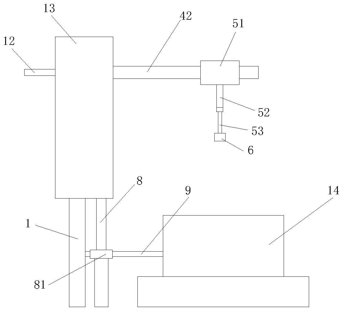
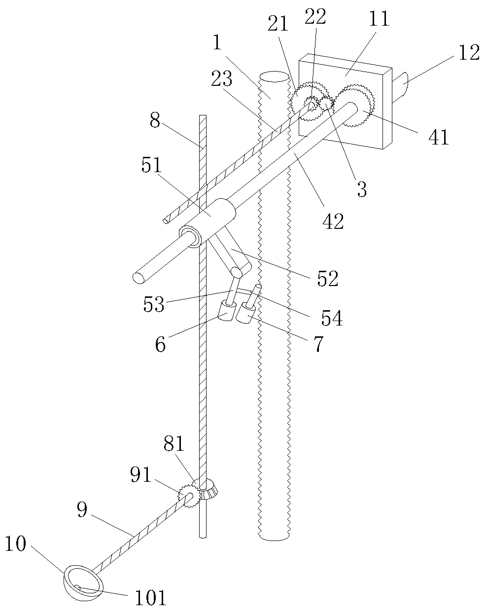
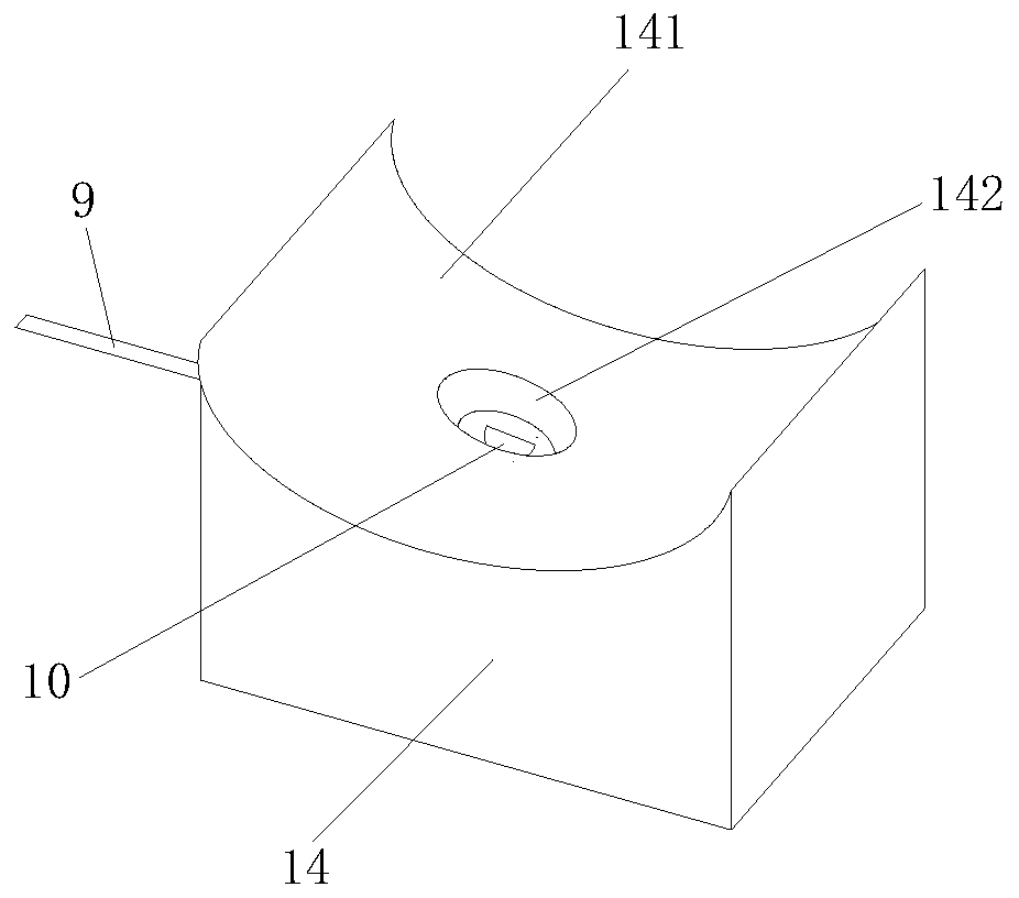
[0047] Example 1. A vascular imaging instrument, such as Figure 1-5 As shown, it includes a vertical rod 1, a gear set, a connecting rod three 8, a connecting rod four 9, a base 14 and a housing 13.

[0048] The gear set includes gear one 21, gear two 22, gear three 3 and gear four 41.

[0049] Gear one 21, gear two 22, gear three 3, and gear four 41 are all installed on the connecting plate 11, and the connecting plate 11 is fixedly provided with a push rod 12, which can move the push rod 12 vertically.

[0050] The vertical rod 1 is arranged vertically, and the outer surface of the vertical rod 1 is provided with a plurality of gear teeth along the vertical direction. The vertical rod 1 is engaged with the gear 21, and the gear 21 can roll up and down along the outer surface of the vertical rod 1. By pushing the push rod 12 up and down, the gear 21 can roll up and down along the outer surface of the vertical rod 1.

[0051] Gear one 21 and gear two 22 are sequentially mounted on c...


PUM


Abstract

The invention relates to the field of medical instruments and particularly relates to a blood vessel development instrument. The blood vessel development instrument comprises an upright rod, a gear set, a connecting rod III, a connecting rod IV and a base, wherein the gear set comprises a gear I, a gear II, a gear III and a gear IV, a gear is arranged on the external surface of the upright rod, the gear I is engaged with the external surface of the upright rod, the gear I can roll up and down along the external surface of the upright rod, the gear I and the gear II are sequentially arranged ona connecting rod I, the gear II, the gear III and the gear IV are engaged with one another sequentially, the gear IV is mounted on a connecting rod II, and a circular ring sleeves the connecting rodII. The invention further provides a use method for the blood vessel development instrument. A snoot and a camera are the same in angle and direction of turning, and the camera is always over againstthe snoot. The blood vessel development instrument disclosed by the invention is simple in use and diverse in function; and through matched use of the gear set and bevel gears, the camera and the snoot can turn in an equidirectional manner during up-and-down drawing of a pusher bar.

Description

Technical field [0001] The invention relates to the field of medical appliances, in particular to a blood vessel imaging instrument and a method of use thereof. Background technique [0002] Venous puncture is one of the most basic and most widely clinically used nursing technical operations in the medical industry. Intravenous injection has also become an important route of administration for clinical treatment and rescue of patients. However, because the blood vessels of some special populations are not easy to find, such as children, obese people, elderly people, long-term intravenous people, people with darker skin, etc., it is rare to have a successful injection, which will increase the pain and increase the patient’s pain. The psychological pressure of nurses can easily lead to medical disputes. Therefore, it is necessary to use a vascular imaging device, which can find the venous blood vessels hidden under the skin and cannot be observed with the naked eye through infrare...

Claims


Application Information

Patent Timeline
Patent Type & Authority Applications(China)
IPC IPC(8): A61M5/42
CPCA61M5/427
Inventor 黎建军
Owner 杭州杭睿科技有限公司
Learn More