
Shear wave imaging method and system

An imaging method and shear wave technology, applied in ultrasonic/sonic/infrasonic Permian technology, ultrasonic/sonic/infrasonic image/data processing, material analysis using sonic emission technology, etc. Cut wave extraction requires high requirements and data processing load is large, so as to reduce the number of detections, reduce the burden of data processing, and concentrate detection energy.

Active Publication Date: 2019-11-29
SHENZHEN MINDRAY BIO MEDICAL ELECTRONICS CO LTD
View PDF6 Cites 4 Cited by
  • Summary
  • Abstract
  • Description
  • Claims
  • Application Information

AI Technical Summary

Problems solved by technology

However, in the application of this method, the shear wave generated inside the tissue is relatively weak, and the propagation of the shear wave in the tissue is a transient process, and the shear wave will attenuate and disappear after a certain time and a certain distance, so it must be Rapid and large-area extraction of shear wave information within a period of time requires high requirements for shear wave extraction, and the data processing load of the extraction system for shear wave extraction is relatively large, and the accuracy is low

Method used


Image

  • Shear wave imaging method and system
  • Shear wave imaging method and system
  • Shear wave imaging method and system

Examples


Embodiment Construction

[0043] The following will clearly and completely describe the technical solutions in the embodiments of the present invention with reference to the accompanying drawings in the embodiments of the present invention. Obviously, the described embodiments are only some, not all, embodiments of the present invention. Based on the embodiments of the present invention, all other embodiments obtained by persons of ordinary skill in the art without creative efforts fall within the protection scope of the present invention.

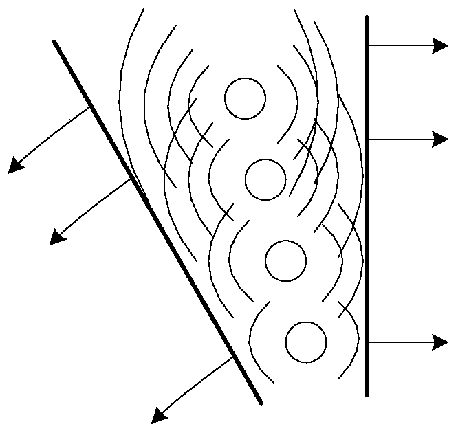
[0044] see figure 1 A preferred embodiment of the present invention provides a shear wave imaging method, which predicts the detection position of the shear wave in advance, so that the detection of the shear wave can be accurately carried out in a small range, so that the detection energy is relatively concentrated, and the detection signal to noise is improved. Compare. At the same time, the number of redundant detections is reduced, the detection process is acc...


PUM


Abstract

The invention provides a shear wave imaging method. The method comprises the following steps of: generating shear wave inside a tissue; pre-estimating the shear wave, sending a plurality of tracking pulses corresponding to positions of the shear wave at different moments, and receiving echo information of the tracking pulses; carrying out shear wave parameter calculation according to the echo information of the tracking pulses; and imaging and displaying the result of the shear wave parameter calculation. According to the shear wave imaging method and system provided by the invention, the detection positions of the shear wave is estimated in advance, so that the detection of the shear wave can be accurately carried out in a small range, the detection energy is relatively concentrated, andthe detection signal-to-noise ratio is improved. Meanwhile, the redundant detection times are reduced, the detection process is accelerated, and the data processing burden is reduced. The invention also provides a shear wave imaging system.

Description

technical field [0001] The invention relates to the field of ultrasonic imaging, in particular to a shear wave imaging method and system. Background technique [0002] Ultrasound elastography is one of the hotspots of clinical research in recent years. It mainly reflects the elasticity or hardness of tissues. It has been increasingly used in the auxiliary detection of tissue cancer lesions, the distinction between benign and malignant, and the evaluation of prognosis and recovery. An existing ultrasonic elastography uses shear waves to perform imaging, mainly by generating shear wave propagation inside the tissue and detecting its propagation parameters and imaging, thereby reflecting the difference in hardness between tissues. [0003] This method has better stability and repeatability. However, in the application of this method, the shear wave generated inside the tissue is relatively weak, and the propagation of the shear wave in the tissue is a transient process, and th...

Claims


Application Information

Patent Timeline
Patent Type & Authority Applications(China)
IPC IPC(8): A61B8/08
CPCA61B8/08G01N29/14
Inventor 李双双樊睿
Owner SHENZHEN MINDRAY BIO MEDICAL ELECTRONICS CO LTD
Learn More