Method for quantifying tumor immune state based on radiomics

A radiomics and state-of-the-art technology, applied in the field of image processing, can solve problems such as different prognosis, inability to obtain pathological specimens, inability to perform surgical resection, etc., and achieve the effect of precise treatment

Active Publication Date: 2019-10-29
GUANGDONG GENERAL HOSPITAL
View PDF2 Cites 11 Cited by
  • Summary
  • Abstract
  • Description
  • Claims
  • Application Information

AI Technical Summary

Problems solved by technology

However, even though some patients have the same TNM stage and histological type, they have different prognoses, and many patients have advanced tumors that cannot be surgically removed, that is, pathological specimens cannot be obtained.

Method used


Image

  • Method for quantifying tumor immune state based on radiomics
  • Method for quantifying tumor immune state based on radiomics
  • Method for quantifying tumor immune state based on radiomics

Examples


Embodiment

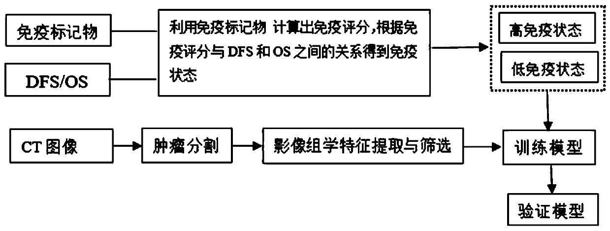
[0034] The sample (colorectal cancer) used in the embodiment of the present invention came from Guangdong Provincial People's Hospital (February 2006-February 2009). This experiment passed the Ethics Committee of Guangdong Provincial People's Hospital, and the patient's informed consent has been obtained.

[0035] The clinical information in this example includes CEA, CA-199, tumor invasion depth, lymph node metastasis, differentiation status, tumor size, Lauren's surgery score, gender, age, TNM stage and overall survival (OS) and disease-free survival (DFS). Among them, CEA and CA-199 were provided by the laboratory department; tumor infiltration depth, TNM and differentiation status were reviewed by attending pathologists; lymph node metastasis and tumor size were reviewed by attending doctors and above.

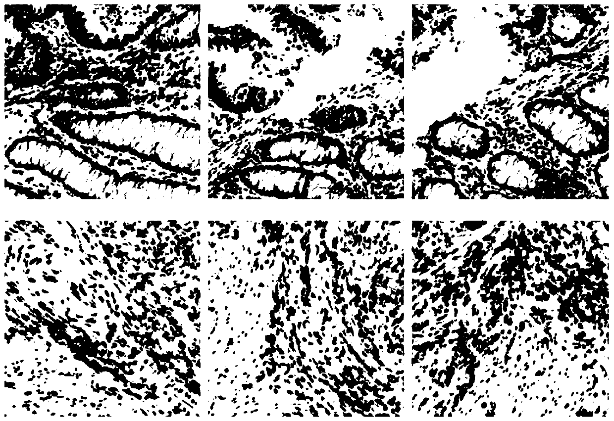
[0036] Such as figure 2 As shown, paraffin tissue slices and baked slices: prepare paraffin-embedded samples into 4 μm serial slices, place the obtained tissue slices on...


PUM


Abstract

The invention discloses a method for quantifying a tumor immune state based on radiomics, wherein the method comprises the following steps: S1, collecting clinical information, immunohistochemical characteristics, overall survival OS and disease-free survival DFS of tumor patients; S2, obtaining a CT image sample group of solid tumors; S3, randomly dividing the patients into a training set and a test set in proportion; S4, calculating the immunoscore by an immune marker, and dividing the patients into a set with high immune state and a set with low immune state; S5, segmenting an interest region, and extracting radiomics characteristics of the solid tumors in the interest region; S6, screening the radiomics characteristics related to the immune state from the radiomics characteristics in the training set, and training a prediction model; and S7, predicting the image immunoscore of the tumor patients, obtaining the image immune state, and then predicting the DFS and the OS in the patients. The immune state of the solid tumors such as colorectal cancer, lung cancer, gastric cancer and breast cancer is predicted by the CT image characteristics, and the analysis of pathological sections and immunohistochemistry is reduced or avoided.

Description

technical field [0001] The invention belongs to the technical field of image processing, and in particular relates to a method for quantifying tumor immune status based on radiomics. Background technique [0002] Currently, biopsy of pathological specimens of tumor patients, pathological staging, and then guiding clinical treatment are commonly used clinical detection methods. However, even though some patients have the same TNM stage and histological type, they have different prognosis, and many patients have advanced tumors when they are found and cannot be surgically removed, that is, pathological specimens cannot be obtained. Therefore, there is an urgent need for a simpler, faster and more accurate method to make up for the deficiency of TNM staging. [0003] Preoperative CT scanning is one of the routine preoperative examination items for patients, and it is applicable to a wide range of people and the examination time is short. Moreover, CT images contain a lot of i...

Claims


Application Information

Patent Timeline
IPC IPC(8): G16H30/20G06T7/00
CPCG06T7/0012G06T2207/10081G06T2207/20081G06T2207/20104G06T2207/30096G16H30/20
Inventor 刘再毅梁长虹揭光灵赵可张芳何兰王瑛陈鑫黄燕琪颜黎栩李振辉
Owner GUANGDONG GENERAL HOSPITAL
Why Eureka
Try Eureka