Device for monitoring intracranial wound healing, its preparation method and application
A wound healing and monitoring device technology, applied in applications, printing devices, diagnostic recording/measurement, etc., can solve the problems of inability to realize real-time imaging of tissue wounds, unavailability of long-term observation, and long time to generate images.
- Summary
- Abstract
- Description
- Claims
- Application Information
AI Technical Summary
Problems solved by technology
Method used
Image
Examples
Embodiment 1
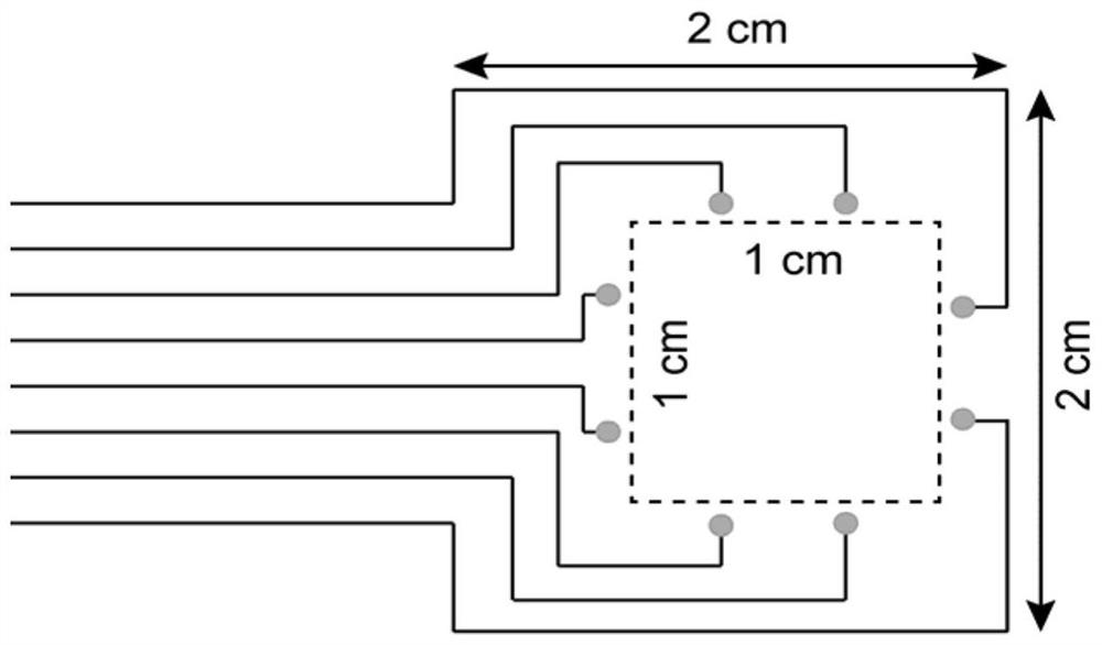
[0045] This embodiment is used to illustrate the processing and preparation method of the flexible brain electrode array of the present invention, which includes the following steps:
[0046] Add 4 grams of liquid metal (indium-gallium alloy, 75.5% gallium mass fraction, 24.5% indium mass fraction) and 1 ml n-decanol into a 5 ml centrifuge tube. The liquid metal micro-nano particles can be prepared by ultrasonicating the ultrasonic cell breaker at 20% intensity for 2 minutes, and the prepared liquid metal ink is added to the screen printing template, and the pattern of the electrode array is printed. Next, cast the pre-cured PDMS (PDMS: curing agent = 10:1) onto the electrode array pattern, put the electrodes in an oven at 80 degrees Celsius to cure for 2 hours, and after the PDMS is completely cured, peel off the PDMS from the substrate A flexible brain electrode array can be obtained. The PDMS is poured on the PET substrate, and after curing is complete, the PDMS is peeled ...
Embodiment 2
[0048] This test example is used to illustrate the flexible electrode array combined with the photoacoustic imaging system to monitor the condition of intracranial wounds, including the following steps:
[0049]Rats are used as experimental subjects, and brain tissue wounds in specific areas of rats are monitored. After the rat was anesthetized, a 2 cm x 2 cm opening was made on the cranium. After the dura was removed, the surface layer was completely exposed. The flexible electrode prepared in Example 1 was attached to the cerebral cortex. The flexible electrode was compatible with the commercial EEG system. connected to monitor changes in EEG signals in the area. At the same time, the photoacoustic imaging system is used to accurately image the monitoring site in real time. The photoacoustic imaging system probe is aimed at the middle area of the brain tissue, the electrode array detects peripheral EEG signals, and the photoacoustic imaging system performs real-time imagin...
PUM
 Abstract
Description
Claims
Application Information
 - R&D
- Intellectual Property
- Life Sciences
- Materials
- Tech Scout
- Unparalleled Data Quality
- Higher Quality Content
- 60% Fewer Hallucinations
Browse by: Latest US Patents, China's latest patents, Technical Efficacy Thesaurus, Application Domain, Technology Topic, Popular Technical Reports.
© 2025 PatSnap. All rights reserved.Legal|Privacy policy|Modern Slavery Act Transparency Statement|Sitemap|About US| Contact US: help@patsnap.com


