endoscopic submucosal dissection mask

An endoscope and lumen technology, applied in the field of endoscopy, can solve the problems of difficult ergonomic control of hinged tools, small working space, difficult simultaneous operation of multiple tools, and obstruction of target tissue visualization.

Active Publication Date: 2021-12-24
BOSTON SCI SCIMED INC
View PDF7 Cites 0 Cited by
  • Summary
  • Abstract
  • Description
  • Claims
  • Application Information

AI Technical Summary

Problems solved by technology

The fully circumferential design of currently available endoscope hoods tends to hinder visualization of target tissue and provides little workspace making it difficult for multiple tools to be manipulated simultaneously
While dual-channel endoscopes may be available in some medical institutions, the separate articulating tools within each channel are often ergonomically difficult to control, especially by a single physician

Method used


Image

  • endoscopic submucosal dissection mask
  • endoscopic submucosal dissection mask
  • endoscopic submucosal dissection mask

Examples


Embodiment Construction

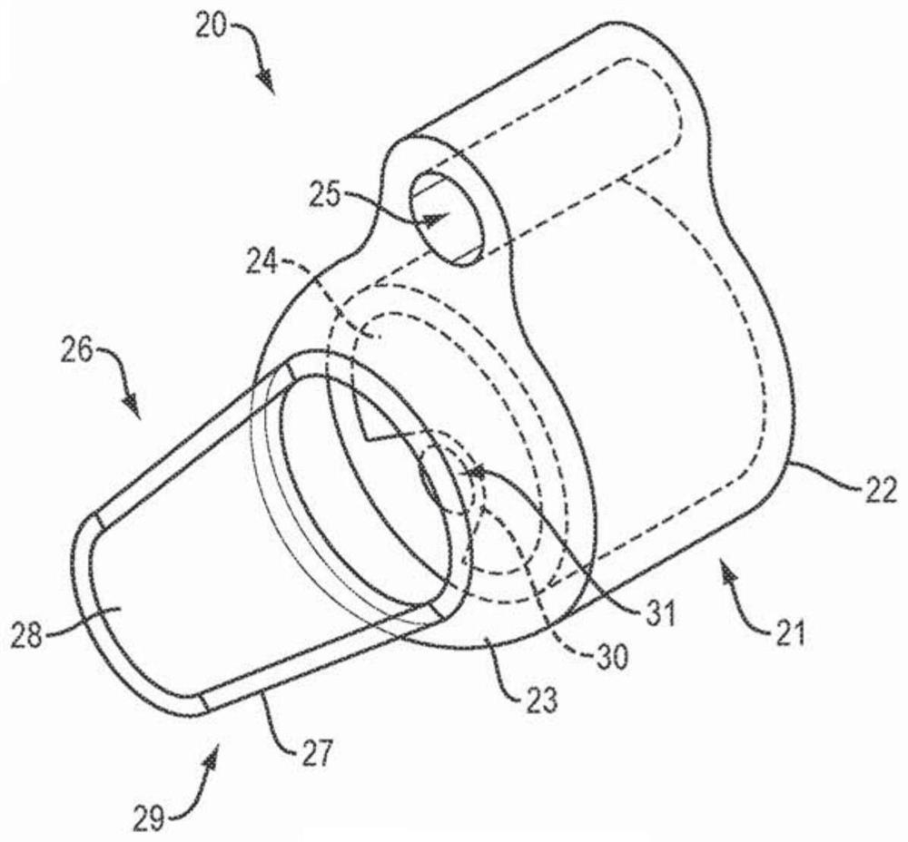
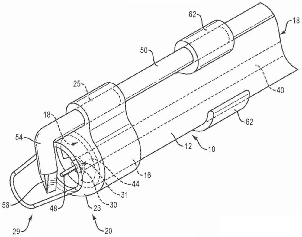
[0018] Before the present disclosure is described in further detail, it is to be understood that this disclosure is not limited to particular embodiments described, as such may vary. It is also to be understood that the terminology used herein is for the purpose of describing particular embodiments only and is not intended to be limiting beyond the scope of the appended claims. Unless otherwise defined, all technical terms used herein have the same meanings as commonly understood by one of ordinary skill in the art to which this disclosure belongs. Finally, although embodiments of the present disclosure are described with particular reference to an endoscope cover attached to the distal end of an endoscope, it should be understood that the endoscope cover disclosed herein may be attached to a Various medical devices including, for example, guide lumens, ports, optical rods, etc. As used herein, the term "distal" refers to the end of the device that is furthest away from the m...


PUM


Abstract

The present disclosure relates to the field of endoscopy. In particular, the present disclosure relates to systems and methods suitable for resecting or dissecting large areas of mucosal or submucosal tissue. The system can apply tension to and continuously manipulate the mucosal tissue such that large lesions can be resected or dissected by a cutting element disposed at the distal end of the endoscope.

Description

[0001] priority [0002] This application claims priority under 35 U.S.C. §119 to U.S. Provisional Patent Application No. 62 / 245,472, filed October 23, 2015, which is hereby incorporated by reference in its entirety for all purposes. technical field [0003] The present disclosure relates to the field of endoscopy. In particular, the present disclosure relates to systems and methods for endoscopic submucosal dissection (ESD) and / or endoscopic mucosal resection (EMR). More specifically, the present disclosure relates to an endoscopic housing that provides enhanced visualization of the working area within which tissue engaging elements and tissue cutting elements operate to dissect or resect mucosal and submucosal lesions. Background technique [0004] Physicians are increasingly willing to perform interventional and therapeutic endoscopic procedures including, for example, full-thickness removal of large tissue lesions such as cancer, submucosal tunneling in the gastrointes...

Claims


Application Information

Patent Timeline
Patent Type & Authority Patents(China)
IPC IPC(8): A61B1/00A61B17/29A61B18/14
CPCA61B17/00234A61B17/29A61B1/00089A61B1/00101A61B18/1492A61B1/00087A61B18/02A61B1/0014A61N7/00A61B17/320016A61B2017/00296A61B2018/00601A61B2017/00269A61B2017/00331A61B2018/1412A61B2018/00982A61B2018/0212A61N2007/0043
Inventor 马修·贾格尔斯基肖恩·赖安赛琳·陈诺曼·梅凯赛琳·伊保尔·史密斯雷·唐
Owner BOSTON SCI SCIMED INC
Why Eureka
Try Eureka