


Quick Research
Generate reliable direction feasibility study reports for your R&D in just a few steps.

Technical Q&A
Discover and master advanced knowledge NOW. Basics, ideas, possibilities, all at once.

Find Solutions
As an expert in R&D theories, this can generate solutions to your technical problems instantly.

Evaluate Feasibility
Analyze your overall solution with one click, know your potential R&D risks in advance.

Monitor Landscape
Get weekly tech updates, stay abreast of the latest tech innovations and key insights.
Breast X-ray photographic system with temperature control function
An X-ray and mammary gland technology, applied in the field of mammography X-ray photography system, can solve the problems of fast heat conduction of the support filter plate and the compression plate, no consideration of the patient’s painful experience, and aggravate the mental burden of the patient, so as to achieve better product design and better product design. Humanization and the effect of improving the patient's medical experience
- Summary
- Abstract
- Description
- Claims
- Application Information
AI Technical Summary
Problems solved by technology
Method used
Image

Examples
Embodiment 1
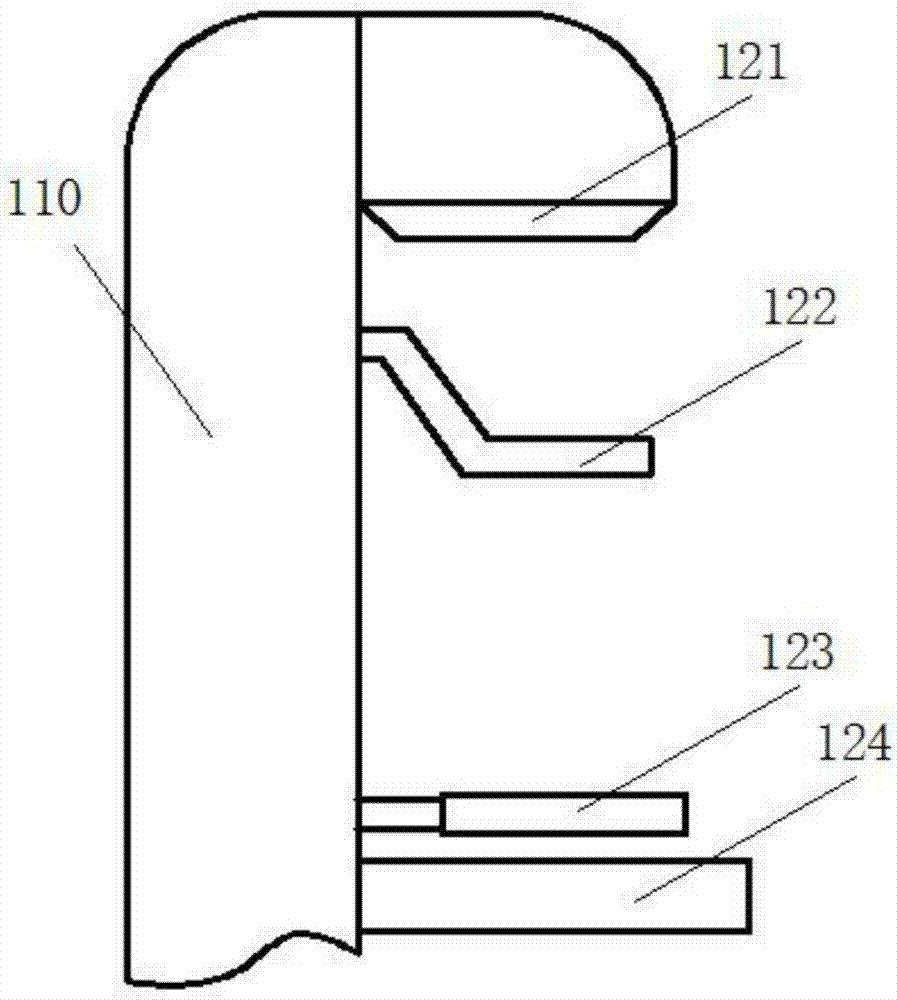
[0028] see figure 1 and figure 2 , as shown in the legend therein, a mammography system includes a main body 100, the main body 100 includes a body 110 and a photographing device installed on the body 10, the photographing device includes a radiation source 121 for irradiating X-rays, A compression plate 122 for compressing the object to be irradiated, a support plate 123 for carrying the object to be irradiated, and an imaging plate 124 for outputting images. The compression plate 122 is provided with a first drive device for driving its compression action and a guide for guiding its compression action. The guide mechanism, the temperature of the compression plate 122 and the support plate 123 can be adjusted and set.
[0029] In the above, from the perspective of the patient’s medical experience, starting from the details of the temperature difference between the support plate and the compression plate and the temperature of the human body, by controlling the temperature o...
Embodiment 2
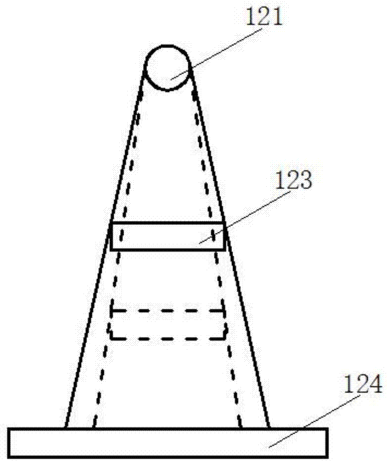
[0038] see image 3 , as shown in the legend therein, the rest are the same as the first embodiment, the difference is that the relative distance between at least one of the ray source 121, the support plate 123 and the imaging plate 124 can be adjusted to make the images relatively The magnification of the irradiated object can be adjusted, the irradiated object is always within the irradiation range of the radiation source 121 , and the image of the irradiated object always falls within the imaging area of the imaging plate 124 .
[0039] In the above, by setting at least one of the ray source, support plate and imaging plate at an adjustable distance from the rest, the FOV of the system can be changed, and the image resolution can be further improved on the basis of unchanged hardware facilities.
[0040] In this embodiment, the relative distance between at least one of the radiation source 121 and the support plate 123 and the human body can be adjusted. , by setting at...
Embodiment 3
[0046] see Figure 4 , as shown in the legend therein, the rest are the same as the first or second embodiment, the difference is that the main body 100 is a horizontal structure, its upper side is set to its human body side and an examination bed board 210 is provided, and the examination bed board 210 is provided with an inspection table. The hole 211 and the solid part of the examination table 210 are set as the first X-ray shielding layer. The human body lies on the examination table 210 , and the object to be irradiated extends from the inspection hole 211 to between the compression plate 122 and the support plate 123 .
[0047]In the above, by setting the first X-ray shielding layer, the patient’s filming process can be made more efficient, safer, and more convenient. It will not affect the effective ray imaging, but also can absorb the radiation damage of the invalid ray to the patient. The cumbersome protective gear makes the shooting process more efficient.
[0048] ...
PUM
 Abstract
Description
Claims
Application Information
 - R&D Engineer
- R&D Manager
- IP Professional
- Industry Leading Data Capabilities
- Powerful AI technology
- Patent DNA Extraction
Browse by: Latest US Patents, China's latest patents, Technical Efficacy Thesaurus, Application Domain, Technology Topic, Popular Technical Reports.
© 2024 PatSnap. All rights reserved.Legal|Privacy policy|Modern Slavery Act Transparency Statement|Sitemap|About US| Contact US: help@patsnap.com