Brain magnetic detection electrical impedance tomography system
An electrical impedance imaging and magnetic detection technology, which is used in telemetry patient monitoring, diagnostic recording/measurement, medical science, etc., can solve the problems of low spatial resolution and small amount of information in EIT imaging, reduce medical risks, and reduce testing costs. , the effect of low cost
- Summary
- Abstract
- Description
- Claims
- Application Information
AI Technical Summary
Benefits of technology
Problems solved by technology
Method used
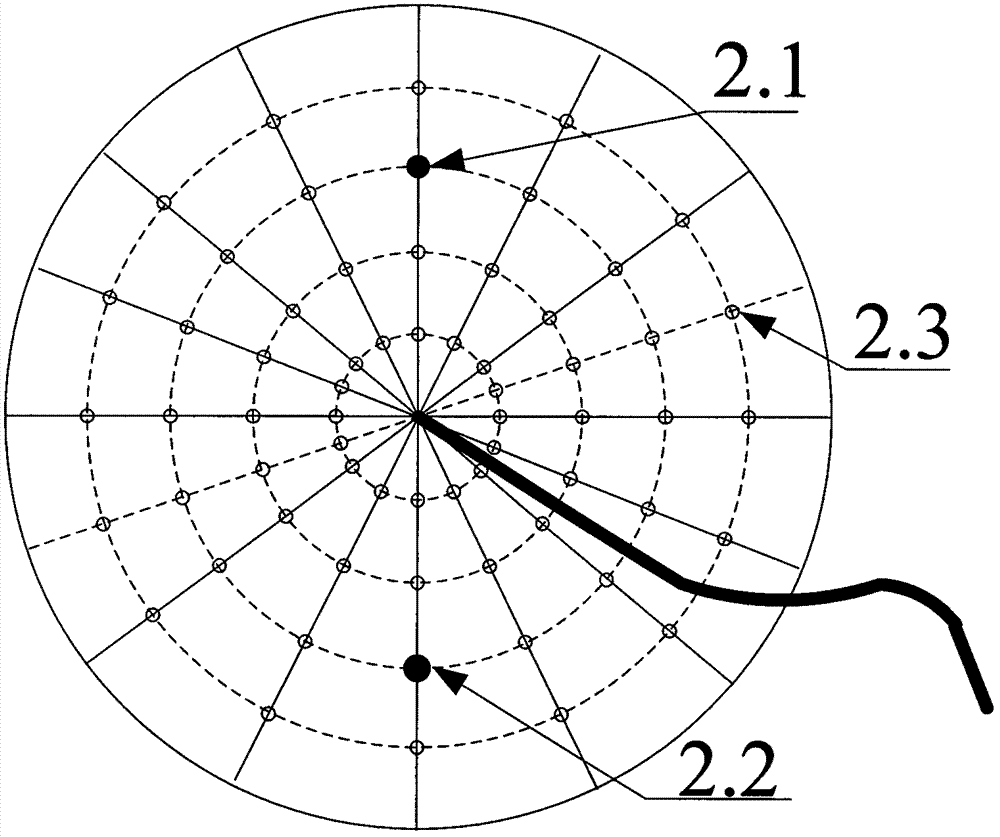
Image


Examples
Embodiment 1
[0047] image 3 It is a schematic diagram of a brain magnetic detection electrical impedance imaging system composed of an electromagnetic detection cap, an excitation acquisition control module, and an image processing display device applied to a static tester. The system usage method is as follows:
[0048] (1) The tester is placed in a supine position.
[0049] (2) Put the electromagnetic detection cap on the tester's head, and inject conductive paste to ensure good contact between the excitation electrode and the skin of the head.
[0050] (3) Apply a certain intensity of safety current to the tester by operating the excitation and acquisition control module, collect the magnetic field distribution around the tester's head in a static state, and transmit the data to the image processing display device.
[0051] (4) The image processing display device uses the image reconstruction algorithm to reconstruct the electrical impedance distribution of the tester's head accordin...
Embodiment 2
[0053] Figure 4 It is a schematic diagram of a tester in motion using a magnetic detection electrical impedance imaging brain diagnostic system composed of an electromagnetic detection cap, an excitation acquisition control module, and a portable image processing display device.
[0054] The system usage method is as follows:
[0055] (1) Put the electromagnetic detection cap firmly on the head of the target to be monitored to ensure good contact between the excitation electrode and the skin of the head.
[0056] (2) Connect the electromagnetic detection cap, the excitation acquisition control module, and the portable image processing display device.
[0057] (3) Apply a certain intensity of safety current to the tester by operating the excitation and acquisition control module, collect the magnetic field distribution around the tester's head in the state of motion, and transmit the data to the image processing display device in real time.
[0058] (4) The image processing ...
PUM
 Abstract
Description
Claims
Application Information
 - Generate Ideas
- Intellectual Property
- Life Sciences
- Materials
- Tech Scout
- Unparalleled Data Quality
- Higher Quality Content
- 60% Fewer Hallucinations
Browse by: Latest US Patents, China's latest patents, Technical Efficacy Thesaurus, Application Domain, Technology Topic, Popular Technical Reports.
© 2025 PatSnap. All rights reserved.Legal|Privacy policy|Modern Slavery Act Transparency Statement|Sitemap|About US| Contact US: help@patsnap.com