
Eye fundus image no-reference quality evaluation method

A fundus image and reference quality technology, applied in image enhancement, image analysis, image data processing, etc., can solve problems such as dark lighting, uneven lighting, and increased medical diagnosis costs

Active Publication Date: 2018-03-30
上海视全视美科技发展有限公司
View PDF3 Cites 23 Cited by
  • Summary
  • Abstract
  • Description
  • Claims
  • Application Information

AI Technical Summary

Problems solved by technology

However, during the imaging process, there are often problems such as brighter lighting, darker lighting, uneven lighting, blurring, low contrast, and unreasonable layout, which lead to the acquisition of fundus images that cannot be used for diagnosis and need to be re-shot, which greatly reduces efficiency and increase the cost of medical diagnosis

Method used


Image

  • Eye fundus image no-reference quality evaluation method

Examples


Embodiment Construction

[0036] The present invention will be further described in detail below in conjunction with the accompanying drawings and embodiments.

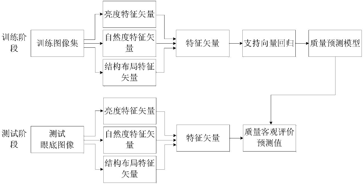
[0037] A no-reference quality evaluation method for fundus images proposed by the present invention, its overall realization block diagram is as follows figure 1 As shown, it includes two processes of training phase and testing phase.

[0038] The specific steps of the described training phase process are:

[0039] ①_1. Select N fundus images to form a training image set, denoted as {I k |1≤k≤N}; Among them, N is a positive integer, N>1, such as taking N=1000, k is a positive integer, 1≤k≤N, I k means {I k |1≤k≤N} in the kth fundus image, {I k The width of each fundus image in |1≤k≤N} is W and the height is H.

[0040] In this embodiment, a part of fundus images in the fundus image database established by Ningbo University is randomly selected to form a training image set.

[0041] ①_2, calculate {I k The brightness feature vector of ea...


PUM


Abstract

The invention discloses an eye fundus image no-reference quality evaluation method. The method comprises two processes including a training stage and a test stage. The influence of brightness, naturalness and structure layout on eye fundus image quality is considered. A dark channel proportion feature, a bright channel proportion feature, a non-uniform brightness feature, a naturalness quality evaluation score and a structure layout index are extracted to form eigenvectors; and then the eigenvectors of all eye fundus images in a training image set are trained by utilizing support vector regression, and a quality prediction model is built. In the test stage, the eigenvectors of the eye fundus images used for test are calculated, and according to the quality prediction model built in the training stage, prediction is performed to obtain quality objective evaluation prediction values of the eye fundus images. The obtained eigenvector information can better reflect the quality change conditions of the eye fundus images, so that the correlation between objective evaluation results and subjective perception is effectively improved.

Description

technical field [0001] The invention relates to an image quality evaluation method, in particular to a no-reference quality evaluation method for fundus images. Background technique [0002] The fundus image is captured by a special fundus camera. The fundus image includes the main physiological structures such as the optic disc, macula, and blood vessels in the retina. It is an important type of image in medical imaging. Among them, the optic disc appears as a nearly circular bright color area in the normal fundus image, which has the strongest contrast with the background area, and is the initial area of ​​the optic nerve and blood vessels; the macula is rich in lutein, so it is the most prominent in the fundus image. It appears as a dark area, and there is no blood vessel structure in this area, and there is an inward sunken area called the fovea in the center of the macula; the blood vessels start from the optic disc area and extend to the entire interior of the eyeball,...

Claims


Application Information

Patent Timeline
IPC IPC(8): G06T7/00G06K9/46G06K9/62
CPCG06T7/0012G06T2207/30041G06T2207/20081G06V10/40G06F18/214G06F18/2411
Inventor 邵枫杨艳李福翠
Owner 上海视全视美科技发展有限公司
Learn More