
A microvascular stapler

A stapler and microvascular technology, applied in the field of medical devices, can solve the problems of complex structure of vascular stapler, prone to vascular embolism, large residual area, etc., to reduce the risk of vascular embolism, reduce the risk of vascular embolism, and small contact area Effect

Inactive Publication Date: 2020-01-17
王培吉
View PDF10 Cites 2 Cited by
  • Summary
  • Abstract
  • Description
  • Claims
  • Application Information

AI Technical Summary

Problems solved by technology

[0004] The present invention provides a microvascular stapler, which aims to solve the problems of complex structure, large residual area after operation and easy occurrence of vascular embolism in the current vascular stapler.

Method used


Image

  • A microvascular stapler
  • A microvascular stapler
  • A microvascular stapler

Examples


Embodiment

[0023] Embodiment: a kind of microvascular stapler

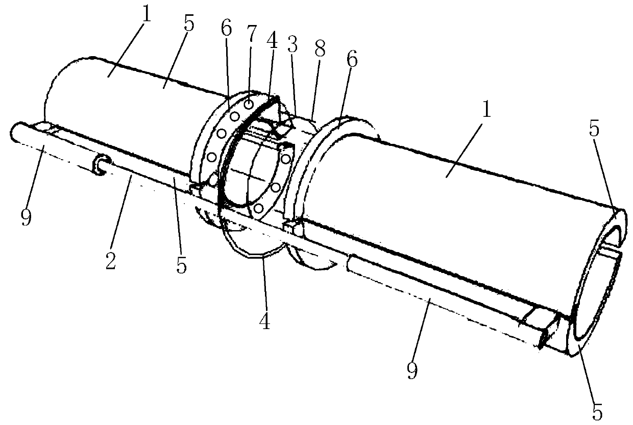
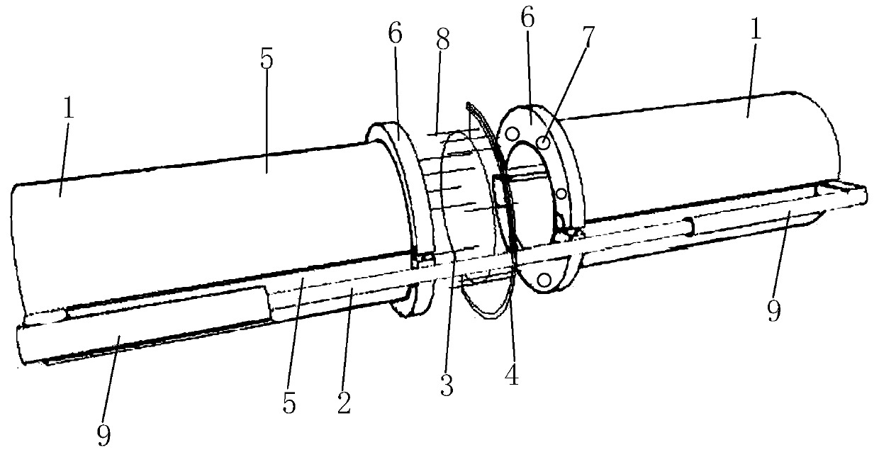
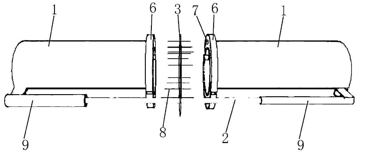
[0024] The microvascular stapler includes two sleeves 1 with the same diameter, a sliding rod 2, staples 3 and a nail seat 4; the sleeve 1 is a tubular member formed by hinged and joined together of two arc-shaped segments 5, The outer periphery of the mouth of one end of the casing 1 is provided with a flange 6, and the flange 6 is provided with a plurality of negative pressure holes 7 along the mouth of the pipe, and the negative pressure holes 7 are specifically through holes; the slide bar 2 is a rod-shaped member, and the two sleeves 1 are slidably connected to the sliding rod 2 on the outer wall of the tube in a symmetrical manner with the flange 6 facing each other. The specific sliding connection method is that the sleeve 1 is fixed with a sliding sleeve 9 , the sliding sleeve 9 is sleeved on the sliding rod 2, so that the sleeve 1 can slide relative to the sliding rod 2, of course, other sliding connection methods i...


PUM


Abstract

Disclosed is a microvascular anastomat. The microvascular anastomat is characterized in that the microvascular anastomat comprises two cannulas same in diameter, a sliding rod, an anastomosis nail anda nail base; each cannula is a tubular member formed by splicing two arc tube sheets in a hinged mode, a flanging arranged at the periphery of a tube opening in one end of each cannula, and a plurality of negative pressure holes are formed in the flangings around the tube opening; the sliding rod is a rod-shaped member, and the outer walls of the two cannulas are both connected with the sliding rod in a symmetrical and sliding mode, wherein the flangings of the two cannulas are opposite to each other; the nail base is a semi-annular member and is fixedly arranged on the sliding rod and located between the two cannulas; the main body of the anastomosis nail is an annular member, the annular member is fixedly provided with a plurality of pins parallel to the central axes of the cannulas, the middle of each pin is fixed to the annular member, and the whole anastomosis nail on the annular member is fixed on the nail base. The microvascular anastomat is simple in structure, the inner membrane eversion of microvessel is maintained after anastomosis, the contact area between the anastomosis nail and the micorvessel after an operation is small, and the risk of postoperative vascular embolism is greatly reduced.

Description

technical field [0001] The invention relates to the field of medical instruments, in particular to a microvessel stapler, which is suitable for anastomosis of microvessels in hand and foot surgery. Background technique [0002] For completely severed fingers, microscopic finger replantation surgery involves the anastomosis and repair of the digital arteries and veins. Finger arteries and veins are microvessels with a diameter of about 1 mm. The usual anastomosis method is manual suturing under a microscope, which requires high suturing skills and is difficult for doctors. The anastomosis effect is difficult to guarantee, and the suture time is long. Usually, it takes about 30 minutes to anastomose a microvessel, which prolongs the microscopic severed finger. Total operating time for implant surgery. Moreover, manual suturing can also damage the intima of the blood vessel, and even cause thrombosis. [0003] At present, there are some vascular staplers on the market, but m...

Claims


Application Information

Patent Timeline
Patent Type & Authority Patents(China)
IPC IPC(8): A61B17/115
Inventor 王培吉崔满意
Owner 王培吉
Learn More