
MRI Image Segmentation Method Based on Fuzzy Thought and Level Set Framework

A level set, fuzzy technology, applied in image analysis, image enhancement, image data processing and other directions, can solve the problem of inability to accurately complete the segmentation process, and the algorithm takes a long time.

Active Publication Date: 2019-01-11
XIDIAN UNIV
View PDF2 Cites 0 Cited by
  • Summary
  • Abstract
  • Description
  • Claims
  • Application Information

AI Technical Summary

Problems solved by technology

However, since the algorithm is improved on the fuzzy clustering algorithm, in each iteration process, the membership matrix u, the cluster center item c, and the update of the local energy item must be completed, and the algorithm takes a long time
And because the fuzzy clustering algorithm has no effective constraint items, the segmentation process cannot be accurately completed in the sharply raised part of the target

Method used


Image

  • MRI Image Segmentation Method Based on Fuzzy Thought and Level Set Framework
  • MRI Image Segmentation Method Based on Fuzzy Thought and Level Set Framework
  • MRI Image Segmentation Method Based on Fuzzy Thought and Level Set Framework

Examples


Embodiment Construction

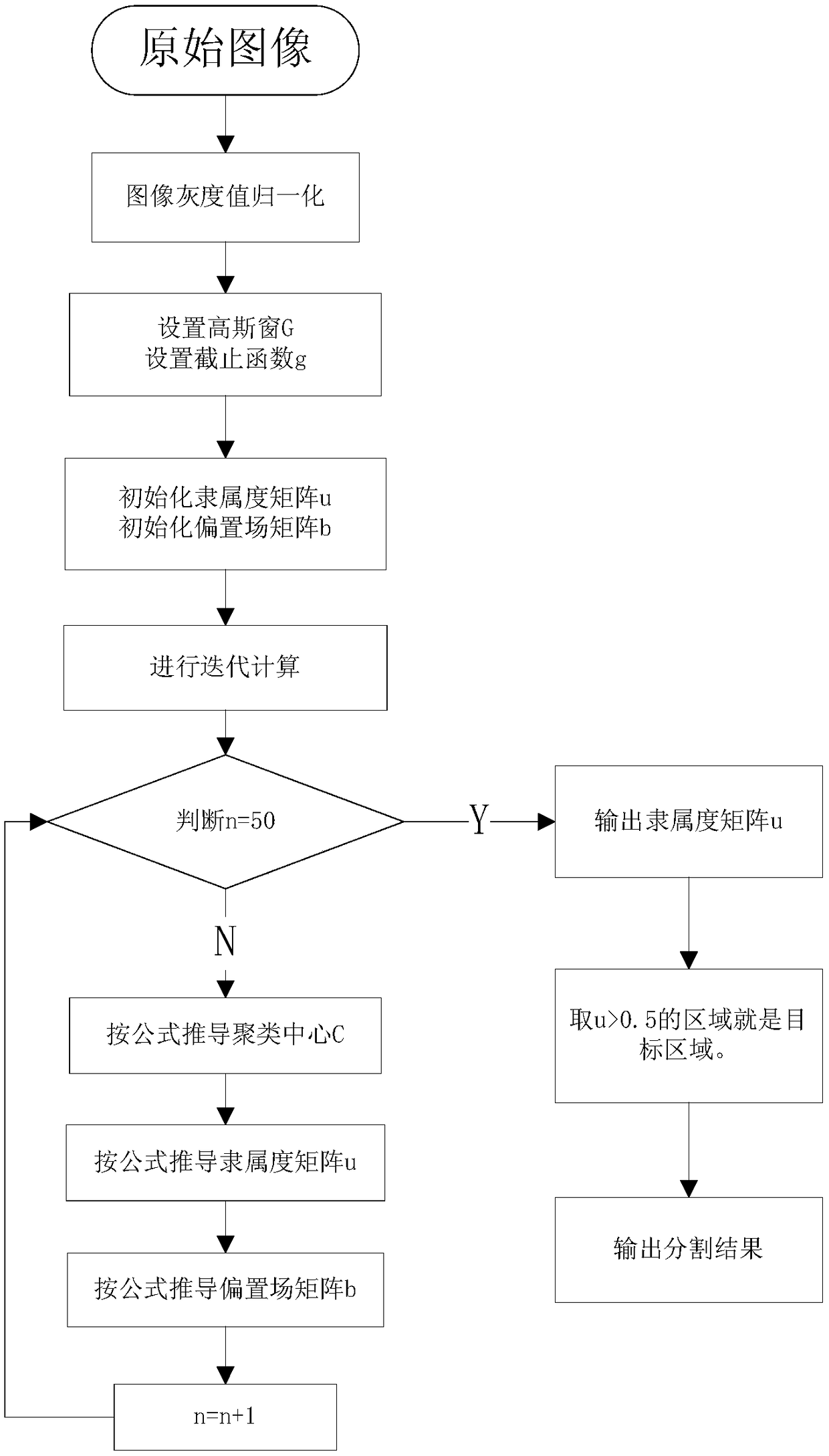
[0035] refer to figure 1 The implementation steps of this example are as follows:

[0036] Step 1 Input brain MRI images.

[0037] Brain MRI image from website http: / / brainweb.bic.mni.mcgill.ca; Take the 40-degree slice MRI image of the brain model as the input original image.

[0038] Step 2 calculates the gray value of each pixel in the image matrix I', and normalizes it. Form a new image matrix I.

[0039] The gray value of all pixels in the brain magnetic resonance image is used to form the image matrix I', and the values ​​in the image matrix I' are normalized to form the nuclear magnetic resonance image matrix I.

[0040] Step 3 Set the initial value of the membership degree matrix u of the brain MRI image:

[0041]

[0042] Where d is a constant item, 00.5, the point belongs to the background area. When u<0.5, the point belongs to the target area. When u =0.5, this point is where the target contour line is located.

[0043] Step 4 sets the initial value of the ...


PUM


Abstract

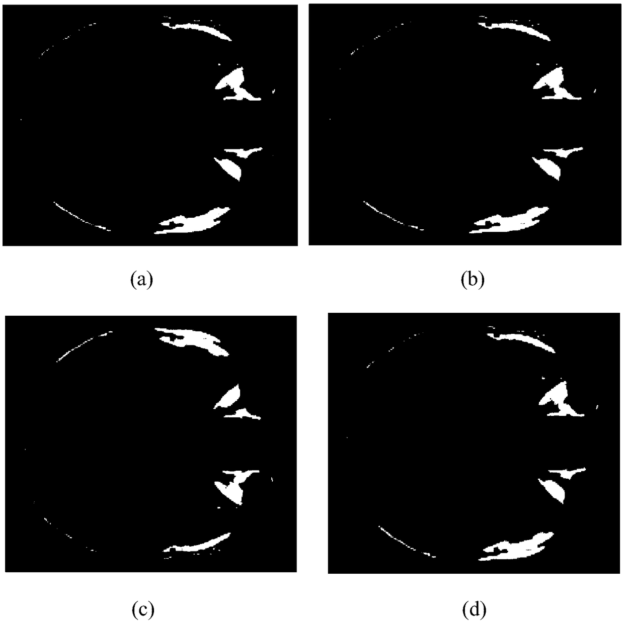
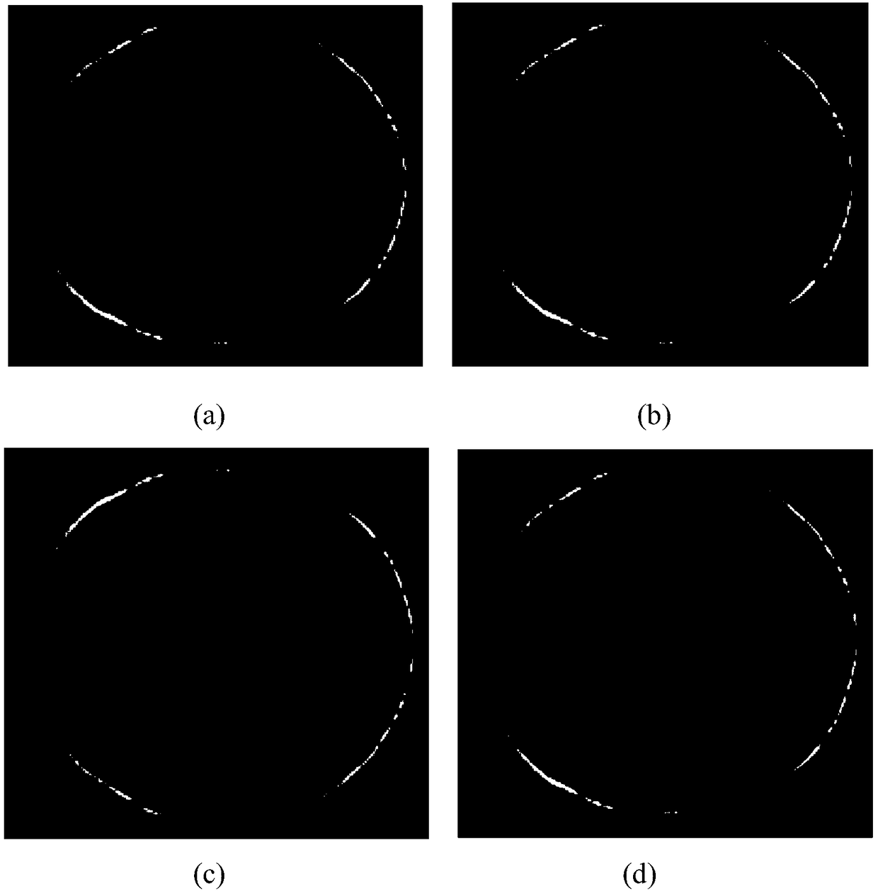
The invention discloses a MRI (Magnetic Resonance Imaging) image segmentation method based on fuzzy thought and a level set frame, and mainly solves the problem of segmentation errors and low calculation speed generated since an original segmentation method can not overcome an intensity non-uniformity phenomenon or noise interference in the MRI process that the nuclear magnetic resonance image is segmented. The method comprises the following implementation steps: 1) inputting an original nuclear magnetic resonance image; 2) carrying out iterative computation on a magnetic resonance image matrix to solve a membership matrix u corresponding to a magnetic resonance image matrix I; 3) regulating an evolution propulsive parameter, and guaranteeing that the membership matrix u is subjected to exact convergence on a target outline; and 4) representing different areas in the image matrix I with different element values of the membership matrix u to obtain an anticipatory segmentation result. By use of the method, the segmentation process of the nuclear magnetic resonance image can be effectively and quickly finished, an accurate target area can be obtained, and the method can be used for cerebral tumor detection and cancer cell examination in a MRI medical image.

Description

technical field [0001] The invention belongs to the field of image information processing and relates to an MRI segmentation method of nuclear magnetic resonance images, which can be used for brain tumor detection and cancer cell inspection in MRI medical images. Background technique [0002] Image segmentation refers to the use of special algorithms to mark out areas of interest to humans in the original image, providing a data basis for feature extraction, recognition, classification, and tracking of image targets. Due to the extensive use of MRI images in medical treatment. Moreover, the inherent non-uniformity of pixel intensity in this type of image interferes with traditional image segmentation algorithms. This makes it impossible for traditional algorithms to draw accurate segmentation conclusions. Therefore, it is of great significance to study the improved segmentation algorithm for this kind of image. In recent years, a large number of scholars have proposed new...

Claims


Application Information

Patent Timeline
Patent Type & Authority Patents(China)
IPC IPC(8): G06T7/10
CPCG06T2207/10088
Inventor 刘靳赵航姬红兵阿鹏仁袁勇董含
Owner XIDIAN UNIV
Learn More