Method and ultrasound apparatus for marking tumor on ultrasound elastography image
A technology of elastic imaging and ultrasonic equipment, which is applied in the directions of ultrasonic/sonic/infrasonic equipment control, ultrasonic/sonic/infrasonic image/data processing, ultrasonic/sonic/infrasonic diagnosis, etc., which can solve the problem that pressure cannot be applied evenly, etc. question
Active Publication Date: 2016-06-29
SAMSUNG ELECTRONICS CO LTD
View PDF5 Cites 6 Cited by
- Summary
- Abstract
- Description
- Claims
- Application Information
AI Technical Summary
Problems solved by technology
However, the freehand elastography method has the disa
Method used
View moreImage
Smart ImageViewing Examples

Examples
 PUM
 Abstract
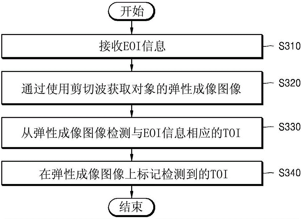
An elasticity information providing method performed by an ultrasound apparatus includes acquiring an elastography image of an object, detecting a region corresponding to a predetermined elastic modulus in the elastography image, and providing information about the region that is detected along with the elastography image.
Description
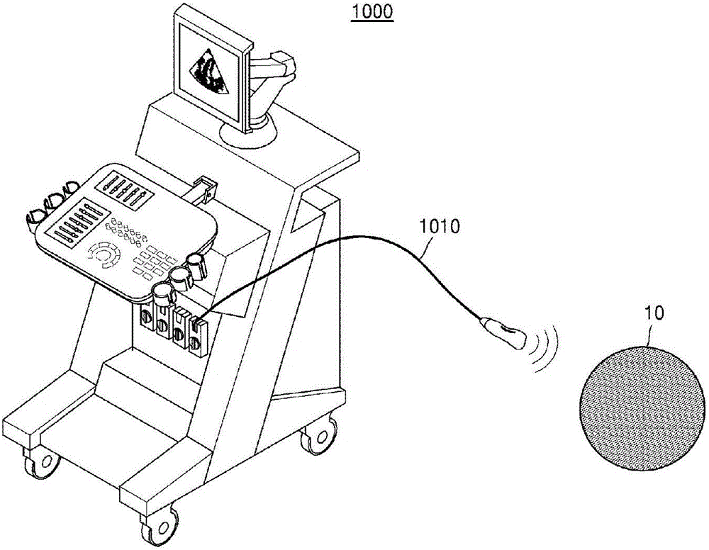
technical field [0001] One or more embodiments relate to a method and ultrasound apparatus for marking a tumor on an elastography image acquired by using shear waves. Background technique [0002] The ultrasonic diagnostic apparatus transmits ultrasonic signals from the surface of a subject to a specific part of a human body, and acquires a tomographic image of soft tissues or an image of blood flow by using information of the ultrasonic signals reflected from internal tissues of the human body. [0003] Ultrasonic diagnostic equipment is small and inexpensive, and displays acquired images in real time. In addition, ultrasonic diagnostic equipment has high stability because the subject is not exposed to X-rays and the like, so ultrasonic diagnostic equipment is different from other image diagnostic equipment such as X-ray diagnostic equipment, computed tomography (CT) scanners, magnetic resonance Imaging (MRI) equipment is widely used together with nuclear medicine diagnost...
Claims
Login to View More Application Information
Patent Timeline
 IPC IPC(8): A61B8/08
CPCA61B8/085A61B8/4405A61B8/4444A61B8/461A61B8/463A61B8/465A61B8/468A61B8/485A61B8/5207A61B8/5223A61B8/54A61B8/00A61B8/08
Inventor 崔基浣朴志瑛孔栋建朴俊浩李炯机
Owner SAMSUNG ELECTRONICS CO LTD
Features
- Generate Ideas
- Intellectual Property
- Life Sciences
- Materials
- Tech Scout
- Unparalleled Data Quality
- Higher Quality Content
- 60% Fewer Hallucinations
Learn More Browse by: Latest US Patents, China's latest patents, Technical Efficacy Thesaurus, Application Domain, Technology Topic, Popular Technical Reports.
© 2025 PatSnap. All rights reserved.Legal|Privacy policy|Modern Slavery Act Transparency Statement|Sitemap|About US| Contact US: help@patsnap.com