Method for detecting vascularization degree of surface of filtering bleb based on ophthalmic slit lamp photographing

A detection method and technology for filtering bubbles, applied in the field of ophthalmic imaging, can solve the problems of difficulty in wide application, expensive instruments, and time-consuming inspection.

Active Publication Date: 2016-05-18
THE EYE HOSPITAL OF WENZHOU MEDICAL UNIV
View PDF6 Cites 7 Cited by
  • Summary
  • Abstract
  • Description
  • Claims
  • Application Information

AI Technical Summary

Problems solved by technology

However, these instruments are relatively expensive, the examination is relatively time-consuming, and some examinations require some invasive o...

Method used


Image

  • Method for detecting vascularization degree of surface of filtering bleb based on ophthalmic slit lamp photographing
  • Method for detecting vascularization degree of surface of filtering bleb based on ophthalmic slit lamp photographing
  • Method for detecting vascularization degree of surface of filtering bleb based on ophthalmic slit lamp photographing

Examples


Embodiment Construction

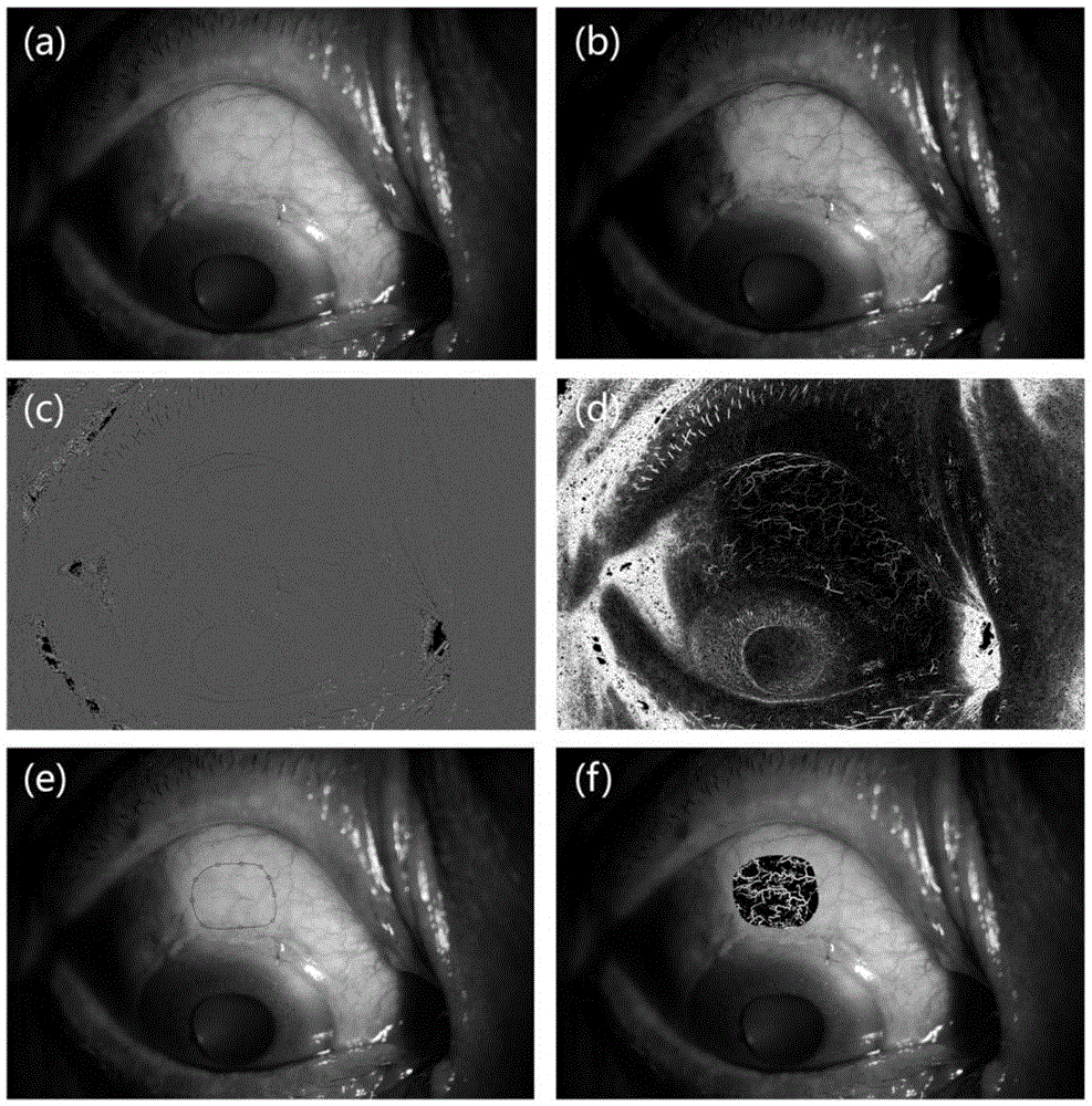
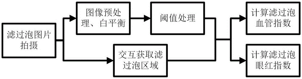
[0032] Based on the slit lamp microscope photography system widely used in clinic, the present invention proposes a detection method of the degree of vascularization on the surface of the filtering bleb based on ophthalmic slit lamp photography. It includes the following steps:

[0033] 1. Collection of filtering bleb images: the patient takes pictures in a dark room. Sit comfortably in front of the slit lamp, place the chin and forehead in front of the bracket, and then the operator gently lifts the patient's upper eyelid, avoiding any pressure on the eyeball, and instructs the patient to look directly downward at the external fixed visual mark, so that the upper eyelid Filtration blebs are fully exposed. The filter bleb was irradiated and photographed under 10 times magnification, 2 levels of light intensity, and diffuse light. It was required to have a panoramic view of the filter bleb, clear blood vessels, and set a control standard white paper below the temporal.

[003...


PUM


Abstract

The invention provides a method for detecting a vascularization degree of the surface of a filtering bleb based on ophthalmic slit lamp photographing. Based on a slit lamp microscope photographing system wide in clinic application, quantification indexes for evaluating blood vessels on the surface of the filtering bleb are provided, the degree of the blood vessels on the surface of the filtering bleb is reflected by a filtering bleb blood vessel area index and a filtering bleb hyperemia index, a vascularization degree objective evaluation method for the surface of filtering bleb is established, and the inflammation state of the filtering bleb is reflected.

Description

technical field [0001] The invention relates to the field of ophthalmology imaging, in particular to a method for detecting the degree of vascularization on the surface of a filtering bleb based on ophthalmic slit lamp photography. Background technique [0002] Trabeculectomy has been used in the treatment of glaucoma since the 1960s. Because it can effectively control intraocular pressure and has a high success rate, it is still the most commonly used external filtration surgery. Postoperative intraocular pressure control depends on the establishment and long-term maintenance of functional aqueous humor drainage channels. The aqueous humor drainage channel includes both the filtering bleb under the conjunctiva, that is, the outer opening of the aqueous humor drainage channel, and the inner opening of the aqueous humor drainage channel under the shallow scleral flap. As an important part of the drainage channel of aqueous humor, the filtering bleb plays a crucial role in th...

Claims


Application Information

Patent Timeline
IPC IPC(8): G06T7/00G06T5/00A61B3/135
CPCA61B3/102A61B3/135G06T5/00G06T5/20G06T7/0012G06T2207/10024G06T2207/10101G06T2207/20032G06T2207/20036G06T2207/20104G06T2207/30041
Inventor 梁远波黄胜海解彦茜
Owner THE EYE HOSPITAL OF WENZHOU MEDICAL UNIV
Why Eureka
Try Eureka