Device and method for X-ray phase contrast imaging

一种X射线、相衬的技术,应用在用于放射诊断的仪器、应用、医药科学等方向,能够解决昂贵等问题

Active Publication Date: 2016-05-04
SIEMENS HEALTHCARE GMBH
View PDF7 Cites 7 Cited by
  • Summary
  • Abstract
  • Description
  • Claims
  • Application Information

AI Technical Summary

Problems solved by technology

Here, however, three X-ray gratings must also be shifted for phase-contrast imaging
The option is therefore more expensive

Method used


Image

  • Device and method for X-ray phase contrast imaging
  • Device and method for X-ray phase contrast imaging
  • Device and method for X-ray phase contrast imaging

Examples


Embodiment Construction

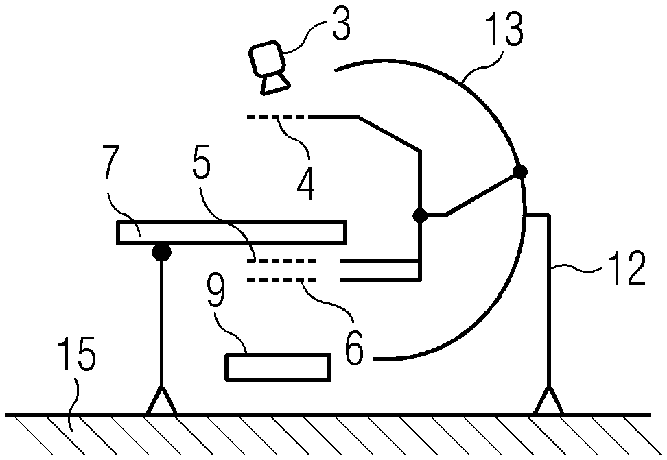
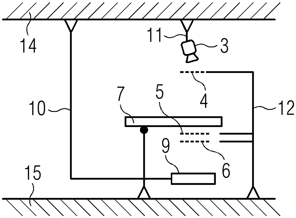
[0035] Figure 1 to Figure 3 An example of a slit scanning device for X-ray phase-contrast imaging with an X-ray emitter 3 , an X-ray detector 9 , a patient bed 7 and a grating arrangement is schematically shown. The grating arrangement has a first x-ray grating 4 , a second x-ray grating 5 and a third x-ray grating 6 . The second and third x-ray grids 5 and 6 are located below the bed board (=operating table) of the hospital bed 7 in front of the x-ray detector 9 . The patient bed 7 is used to accommodate objects (not shown) which are seen through the x-ray fan of the x-ray emitter 3 . The object (for example a patient) can be moved together with the hospital bed 7 .

[0036] exist figure 1 and figure 2 In this case, the X-ray detector 9 is fixed at the first ceiling bracket 10 and can be moved by means of the first ceiling bracket 10 arranged at the ceiling 14 . exist figure 1 , the X-ray radiator 3 is connected to a second ceiling support 11 and can be moved by means...


PUM


Abstract

A device for slot scanning phase contrast X-ray imaging, includes an X-ray emitter (3), a plurality of X-ray gratings (4, 5, 6), a patient couch (7), and an X-ray detector (9). Position marker elements (1, 2) are arranged and configured in the beam path of the X-ray emitter (3) such that the position marker elements (1, 2) are visible in an X-ray image. In the X-ray image, a relative position of an object on the patient couch (7) in relation to an X-ray beam fan of the X-ray emitter (3) may be established from a location and a distance of the position marker elements (1, 2) from one another. The method thereof is illustrated in the same way. The device and the method are advantageous in that there is no need to perform high-cost and accurate mechanical adjustment of assemblies of equipment.

Description

technical field [0001] The present invention relates to equipment and methods for slit scanning (Slot-Scan), X-ray phase contrast imaging. Background technique [0002] X-ray phase-contrast imaging is a new type of X-ray imaging method which does not use only the absorption by the object as a source of information, as in conventional X-ray systems. This method combines absorption with a shift in the phase of the x-ray emission as it passes through the object. The information content is much higher, since on the one hand the absorption provides a precise image of the highly absorbing bone, and on the other hand the phase contrast produces a sharp image of the soft tissue structure. This offers the possibility that pathological changes, such as the development of masses, narrowing of blood vessels or pathological changes in cartilage, can be detected much earlier than before. [0003] The passage of X-ray radiation through a material is described by means of the complex refr...

Claims


Application Information

Patent Timeline
Patent Type & Authority Applications(China)
IPC IPC(8): A61B6/00
CPCA61B6/484A61B6/0407A61B6/0492A61B6/4035A61B6/4291A61B6/5235
Inventor J.弗罗伊登贝格尔
Owner SIEMENS HEALTHCARE GMBH
Why Eureka
Try Eureka