Registration method applied to retina fundus image

A fundus image and retina technology, which is applied in the field of medical image processing, can solve the problems such as the inability to realize retinal fundus images, achieve the improvement of the number of registration points and the registration accuracy, improve the accuracy, and achieve the effect of registration

Inactive Publication Date: 2015-09-23
SHANDONG UNIV
View PDF6 Cites 14 Cited by
  • Summary
  • Abstract
  • Description
  • Claims
  • Application Information

AI Technical Summary

Problems solved by technology

[0004] Aiming at the registration defects that the classic SIFT method cannot achieve in some retinal fundus images, the present invention designs a registration method applied to fundus retinal images

Method used


Image

  • Registration method applied to retina fundus image
  • Registration method applied to retina fundus image
  • Registration method applied to retina fundus image

Examples


Embodiment Construction

[0027] The present invention will be further described in detail below in conjunction with the drawings and specific embodiments.

[0028] The registration process of the medical image in the present invention goes through the following steps in sequence:

[0029] Step 1: Preprocess the two images to be registered to make their types consistent;

[0030] Step 2: Extract the SIFT feature structure point set in the image and perform feature description;

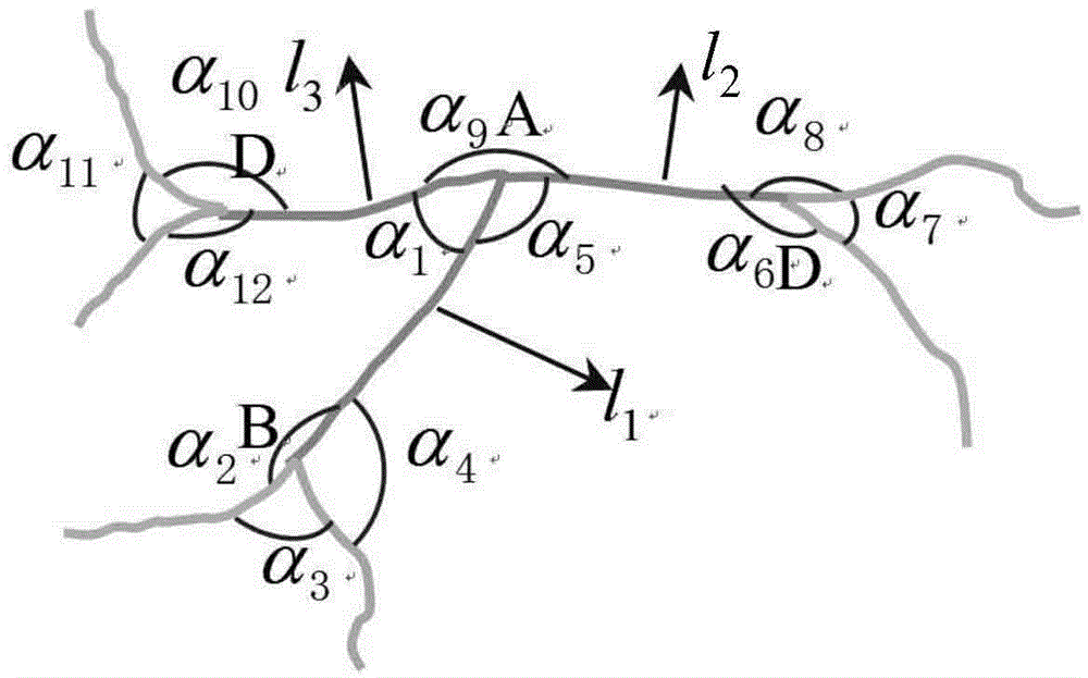
[0031] Step 3: Extract the bifurcation structure point set in the image and perform feature description. The method of feature description is attached figure 1 As shown, a feature structure includes the main bifurcation point and three neighboring nodes around it, k 1 =α 1 / α 5 / α 9 , K 2 =α 2 / α 3 / α 4 , K 3 =α 6 / α 7 / α 8 , K 4 =α 10 / α 11 / α 12 . Right l i And k i To normalize, l i ′ = l i l 1 + l 2 + l 3 , k i ′ = k i ′ k 1 ′ + k 2 ′ + k 3 ...


PUM


Abstract

The invention discloses a registration method applied to a retina fundus image, and belongs to the field of medical image processing. On the basis of an SIFT (Scale Invariant Feature Transform) method, a new feature point detection and feature description method is proposed, the detection amount and the registration precision of the feature points of the retina fundus image can be improved, and an image registration effect is better. In order to further improve registration accuracy, a mismatching fiducial point rejection strategy is introduced into registration. A great quantity of registration experiments indicates that the registration method can realize the accurate and stable registration of the retina fundus image and overcomes a defect that the SIFT method can not realize the registration in parts of retina fundus images.

Description

Technical field [0001] The invention belongs to the field of medical image processing, and particularly relates to a registration method applied to retinal fundus images. Background technique [0002] The retinal fundus image is a standard objective diagnostic image commonly used by ophthalmologists. It is obtained by a fundus camera. Fundus refers to the collective name of the tissue structure of the inner back of the eye, which generally includes the retina, optic nerve, choroid, and macula. Studies have shown that diseases such as glaucoma, diabetic retinopathy and coronary heart disease can cause vision loss, which is the main cause of blindness. In the process of clinical diagnosis and treatment, ophthalmologists need to compare and analyze the retinal fundus images taken by the same patient in different periods. Due to the different imaging angles and imaging environment, high-precision registration of the two images is required at this time. In addition, due to the angl...

Claims


Application Information

Patent Timeline
Patent Type & Authority Applications(China)
IPC IPC(8): G06T7/00
CPCG06T7/0014G06T7/33G06T2207/30041
Inventor 董恩清孙华魁黄振强仲伟冲李宇森
Owner SHANDONG UNIV
Why Eureka
Try Eureka