
Coil arrangement for magnetic resonance tomography equipment

A tomography and magnetic resonance technology, which is applied in magnetic resonance measurement, measurement using nuclear magnetic resonance imaging system, and magnetic variable measurement, etc. Remove simplified, good image quality effects

Inactive Publication Date: 2018-02-27
休伯特·劳拉斯
View PDF4 Cites 0 Cited by
  • Summary
  • Abstract
  • Description
  • Claims
  • Application Information

AI Technical Summary

Problems solved by technology

[0005] However, these receiver coils are relatively far away from the prostate due to their structure
Therefore, only relatively insufficient resolution of the prostate is guaranteed

Method used


Image

  • Coil arrangement for magnetic resonance tomography equipment
  • Coil arrangement for magnetic resonance tomography equipment

Examples


Embodiment Construction

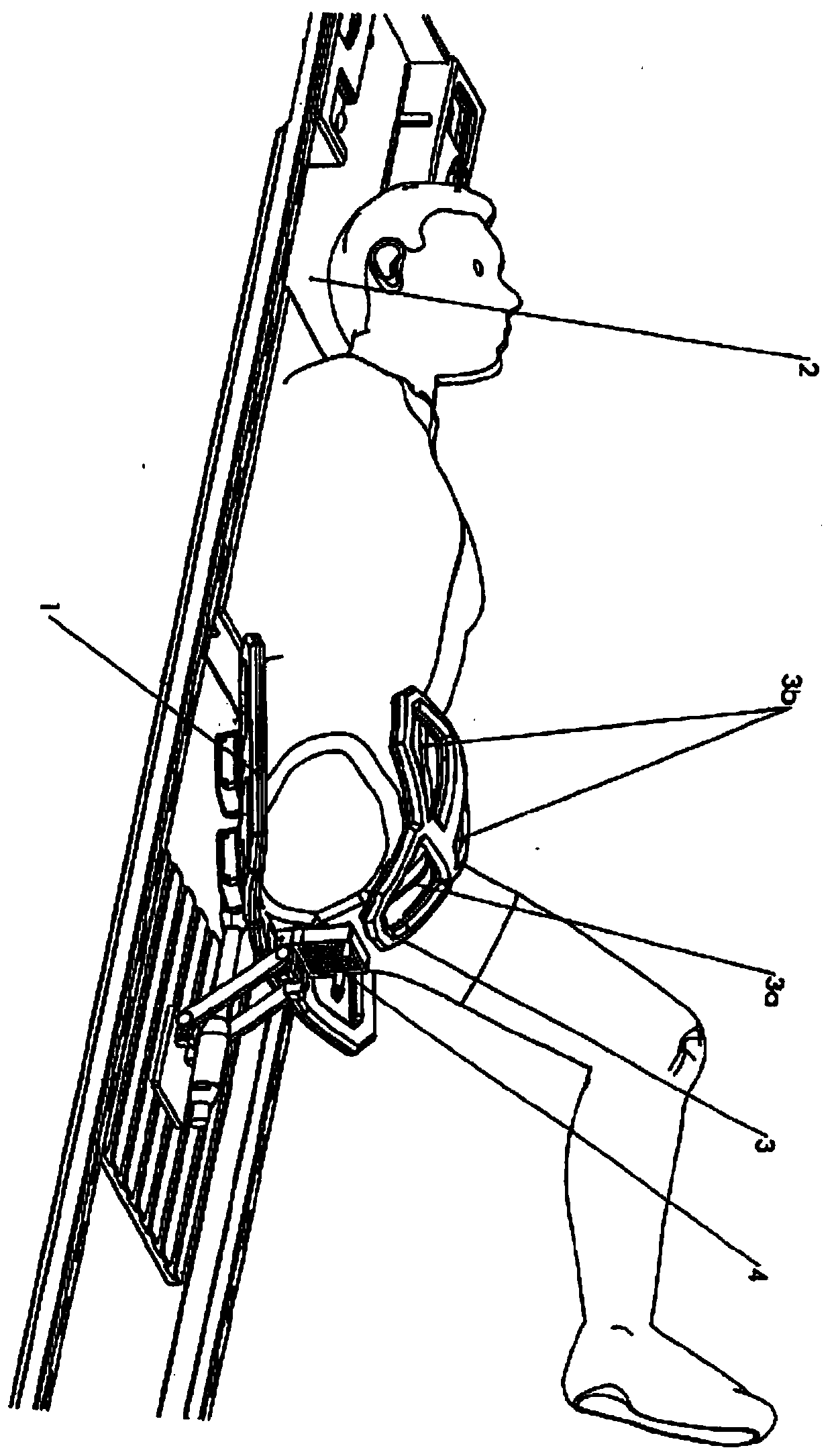
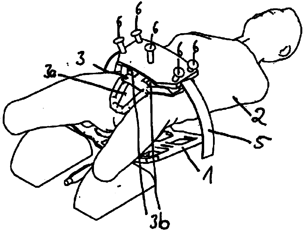
[0028] According to the invention, the coil elements ( 3 ) are positioned on the patient's lower abdomen area and on the groin area. This is divided into three individual coils arranged in a triangle. The two coils ( 3 b ) are positioned next to each other parallel to each other on the patient's lower abdominal region and are oriented with their surface normals in the direction of the prostate. The third coil (3a), which is essential with the present invention, is attached centrally below the two coils (3b) so that it surrounds the scrotum and penis and rests on the patient's groin when the patient stretches his thigh slightly. The third coil

[0029](3a) is V-shaped and is also oriented with its surface normal into the direction of the prostate. The biopsy device ( 4 ) also shown is not functional for the invention. The actual magnetic resonance tomography equipment, that is to say the coils generating the strong homogeneous magnetic field and the transmitter coils, are no...


PUM


Abstract

A magnetic resonance tomography apparatus for performing a prostate examination on a patient in said apparatus, comprising: a coil for generating a strong uniform magnetic field in the direction of the patient's longitudinal axis, at least a transmitting coil, three gradient coils and suitable receiving coils, wherein a single coil is arranged under the patient in the lower back region and / or at the back, and at least one of said coils is arranged above the patient, and also comprising Data processing for obtaining images from the signals of the transmitting and receiving coils, wherein a closed receiving coil is provided in close proximity to the patient and surrounds the scrotum and penis.

Description

technical field [0001] The invention relates to a magnetic resonance tomography apparatus for examining the prostate gland of a patient therein, comprising: a coil for generating a strong uniform magnetic field in the direction of the longitudinal axis of the patient, at least A transmitter coil, three gradient coils and suitable receiver coils, a single coil is arranged under the lower back area and / or on the back and at least one coil is arranged on the patient, and for Signals from the machine coil are processed for imaging data. Background technique [0002] In particular, early identification of prostate cancer is important for successful treatment. Common methods of finding prostate cancer (eg, manual inspection and blood tests) fail to locate certain malignancies or sometimes give false positive test results. A biopsy is the way to locate the tumor and assess its risk. Unfortunately, this biopsy often misses tumors. [0003] Magnetic resonance tomography is an ima...

Claims


Application Information

Patent Timeline
Patent Type & Authority Patents(China)
IPC IPC(8): A61B5/055A61B5/00G01R33/34G01R33/3415
CPCA61B5/055A61B5/4381G01R33/34084G01R33/3415G01R33/30G01R33/385G01R33/54
Inventor 休伯特·劳拉斯
Owner 休伯特·劳拉斯
Learn More