
Image processing method and device for equalizing breast peripheral tissue

An image and tissue technology, applied in the field of medical image processing, can solve problems such as compression amplitude noise, low processing efficiency, and insufficient equalization effect

Active Publication Date: 2017-12-29
NEUSOFT MEDICAL SYST CO LTD
View PDF4 Cites 0 Cited by
  • Summary
  • Abstract
  • Description
  • Claims
  • Application Information

AI Technical Summary

Problems solved by technology

[0002] The distribution characteristics of breast tissue cause a wide range of grayscale differences between the central area and the edge area. If a fixed brightness contrast is used, only a part of the tissue can be observed. If the dynamic range compression method is used, the small compression range will easily cause insufficient equalization effect, and the compression range is too large. It will also cause problems of excessive noise and artifacts, and in severe cases, it may also change the original gray distribution characteristics of the glandular tissue area
[0003] In the prior art, the large-scale convolution kernel method is usually used to equalize the peripheral tissue of the breast. This method specifically uses a fixed compression factor to process the image as a whole. Since the tissue information on the side of the breast wall close to the breast wall is quite different from the density of the peripheral tissue, A fixed compression factor is used. If the compression factor is too large, the original gray features of the gland area may change. If the compression factor is too small, the equalization effect is not enough, and the peripheral tissues cannot be seen clearly. Therefore, a fixed compression factor cannot guarantee that all images in the image In addition, the large-size convolution kernel method often uses a convolution kernel size of 100-300, resulting in a very large amount of calculation for the equalization process and low processing efficiency.

Method used


Image

  • Image processing method and device for equalizing breast peripheral tissue
  • Image processing method and device for equalizing breast peripheral tissue
  • Image processing method and device for equalizing breast peripheral tissue

Examples


Embodiment 1

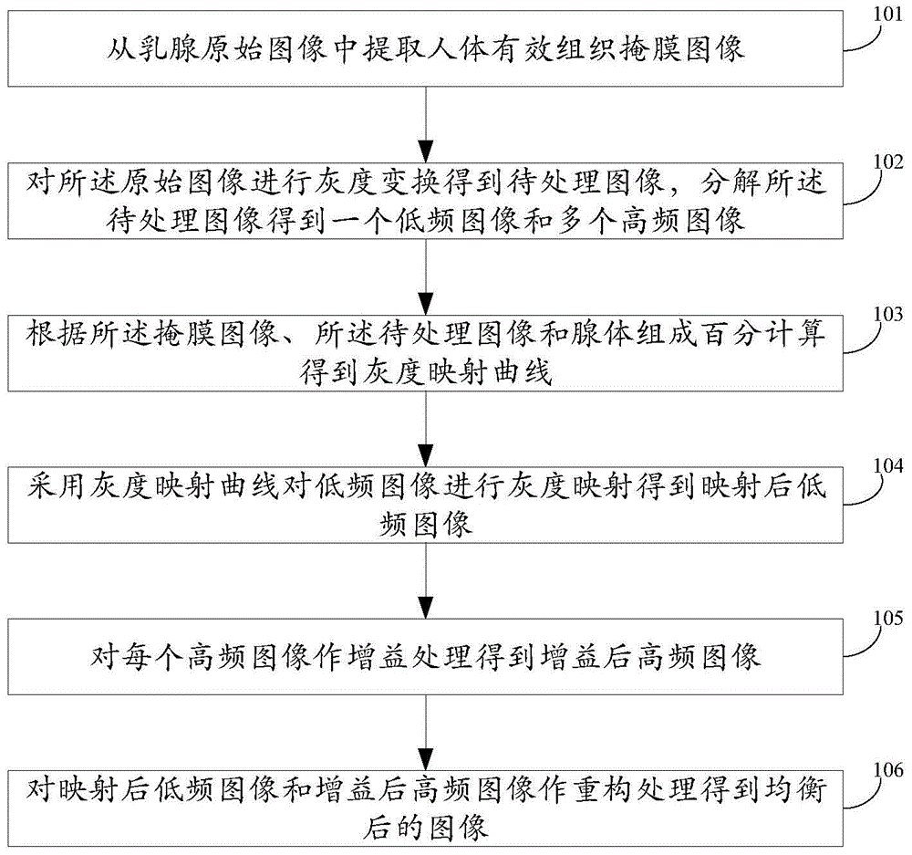
[0070] refer to figure 1 , figure 1 It is a flow chart of an image processing method for equalizing breast peripheral tissue in Embodiment 1 of the present invention. The method in this embodiment may specifically include:

[0071] Step 101, extracting an effective human body tissue mask image from the original image of the mammary gland.

[0072] In this embodiment, firstly, the original image of the mammary gland part is obtained, and secondly, the effective tissue mask image of the human body is extracted from the original image by segmenting the original image. At present, a variety of methods are applicable to the segmentation of the original image of the mammary gland, such as the histogram-based O TSU segmentation method, the method based on region growth and other segmentation methods. Since the original image of the breast portion has a sharp contrast between the human tissue and the background, it is preferred that In the specific implementation of this embodiment,...

Embodiment 2

[0113] refer to figure 2 , figure 2 It is a flowchart of an image processing method for equalizing breast peripheral tissue in Embodiment 2 of the present invention. The method in this embodiment may specifically include:

[0114] Step 201, extracting an effective human body tissue mask image from the original breast image;

[0115] Step 202, performing grayscale transformation on the original image to obtain an image to be processed, and decomposing the image to be processed to obtain a low-frequency image and multiple high-frequency images;

[0116] Step 203, calculating and obtaining a grayscale mapping curve according to the mask image, the image to be processed, and the gland composition percentage;

[0117] Step 204, using the gray-scale mapping curve to perform gray-scale mapping on the low-frequency image to obtain the mapped low-frequency image;

[0118] Step 205, performing gain processing on each high-frequency image to obtain a high-frequency image after gain;...

Embodiment 3

[0129] In order to realize the above method, the present invention also provides an image processing device for balancing breast peripheral tissues.

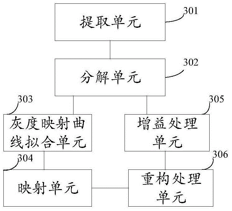
[0130] refer to image 3 , image 3 It is a structural diagram of an image processing device for equalizing breast peripheral tissue in Embodiment 3 of the present invention. The device in this embodiment may specifically include:

[0131] An extraction unit 301, configured to extract an effective tissue mask image of the human body from the original image of the mammary gland;

[0132] Decomposing unit 302, configured to perform grayscale transformation on the original image to obtain an image to be processed, and decompose the image to be processed to obtain a low-frequency image and multiple high-frequency images;

[0133] A grayscale mapping curve fitting unit 303, configured to calculate and obtain a grayscale mapping curve according to the mask image, the image to be processed, and the composition percentage of glands; ...


PUM


Abstract

The embodiment of the invention discloses an image processing method and device for mammary peripheral tissue equalization. The method comprises the following steps: extracting a mask image of effective human tissue from a mammary original image; carrying out grey level transformation on the original image so as to obtain an image to be processed; decomposing the image to be processed so as to obtain a low-frequency image and a plurality of high-frequency images; calculating according to the percentage composition of the mask image, the image to be processed and a gland so as to obtain a grey mapping curve; carrying out grey mapping on the low-frequency image through the grey mapping curve so as to obtain a mapped low-frequency image; carrying out gain processing on all high-frequency images so as to obtain gained high-frequency images; carrying out reconstruction processing on the mapped low-frequency image and the gained high-frequency images, and thus obtaining an equalized image. The method can carry out adaptive adjustment according to individual differences, thereby ensuring the equalization effect, reducing noises and artifacts, and improving the processing efficiency.

Description

technical field [0001] The invention relates to the field of medical image processing, in particular to an image processing method and device for balancing breast peripheral tissues. Background technique [0002] The distribution characteristics of breast tissue cause a wide range of grayscale differences between the central area and the edge area. If a fixed brightness contrast is used, only a part of the tissue can be observed. If the dynamic range compression method is used, the small compression range will easily cause insufficient equalization effect, and the compression range is too large. It will also cause the problem of excessive noise and artifacts, and in severe cases, it may also change the original gray distribution characteristics of the glandular tissue area. [0003] In the prior art, the large-scale convolution kernel method is usually used to equalize the peripheral tissue of the breast. This method specifically uses a fixed compression factor to process th...

Claims


Application Information

Patent Timeline
Patent Type & Authority Patents(China)
IPC IPC(8): G06T7/00G06T7/12G06T7/136G06T7/44
CPCG06T7/0012G06T7/11G06T2207/20048G06T2207/30068
Inventor 李海春
Owner NEUSOFT MEDICAL SYST CO LTD
Learn More