
Lung lobe breath sound monitoring and automatic analyzing device

An automatic analysis device, breath sound technology, applied in medical science, auscultation instrument, diagnosis, etc.

Inactive Publication Date: 2015-04-22
NORTH CHINA UNIVERSITY OF SCIENCE AND TECHNOLOGY
View PDF9 Cites 4 Cited by
  • Summary
  • Abstract
  • Description
  • Claims
  • Application Information

AI Technical Summary

Problems solved by technology

[0003] The purpose of the invention of the present invention is to aim at the low sensitivity of the breathing sound auscultation equipment of the prior art, be affected greatly by man-made or conditional factors, and lack the breath sound of each lung lobe Multi-site, dynamic, intuitive detection and automatic analysis of deficiencies, provide a device that can simultaneously monitor the breath sounds of each lobe of the lungs, and display them in enlarged sound and waveform images. At the same time, the device can automatically analyze breath sounds and waveforms. Store and analyze for medical staff to carry out clinical operations

Method used


Image

  • Lung lobe breath sound monitoring and automatic analyzing device
  • Lung lobe breath sound monitoring and automatic analyzing device

Examples


Embodiment 1

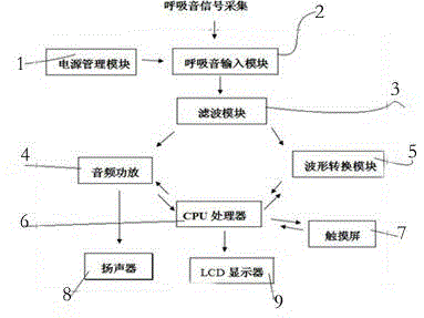
[0024] according to figure 1 Connect the device of the present invention as shown, press figure 2 Connect the breath sound collection device as shown, and turn on the power of the device. The patient had left upper lobe cancer, and planned left lung resection, which required right double-lumen tracheal intubation. Before endotracheal intubation, the LCD monitor 9 displayed breath sound waveforms of 5 lung lobes, and indicated that the breath sounds of the 5 lung lobes were basically normal. Basically normal, indicating that the tracheal intubation has been detected in the trachea; when the left lung is ventilated, L 上 , L 下 The waveform is normal, R 上 , R 中 , R 下 The waveform is obviously weakened or disappeared, the right lung is ventilated, R 上 , R 中 , R 下 The waveform is normal, L 上 , L 下 The waveform is obviously weakened or disappeared, indicating that the double-lumen (right) tracheal intubation is completely successful; if the right lung is ventilated, R ...

Embodiment 2

[0026] according to figure 1 Connect the device of the present invention as shown, press figure 2 Connect the breath sound collection device as shown, and turn on the power of the device. On the day when the patient was admitted to the hospital with bilateral thoracic trauma, lung contusion, and multiple rib fractures, the patient was placed in a semi-recumbent position, and was given ECG monitoring and blood oxygen saturation detection. The breath sound waveforms in each lung lobe were basically normal; on the second day, LCD monitor 9 displayed R 上 , L 上 , Breath sound waveform is basically normal, normal alveolar breath sound, R 下 , L 下The breath sound waveform is abnormal, which is wet rale, suggesting exudative changes in the lower lobes of both lungs; R 中 The waveform of breath sounds was significantly weakened, suggesting that the middle lobe is poorly ventilated. On the third day, the monitoring showed R 上 , L 上 , Breath sound waveform is basically normal, no...


PUM


Abstract

The invention relates to a lung lobe breath sound monitoring and automatic analyzing device which comprises a breath sound signal collecting system, a power supply management module, a breath sound input module, a filter module, an audio power amplifier, a waveform conversion module, a loudspeaker, a CPU processor, a touch screen and an LCD display, wherein the power supply management module is connected with the input end of the breath sound input module by virtue of a wire; the output end of the breath sound input module is connected with the filter module by virtue of a wire; the output end of the filter module is connected with the input ends of the audio power amplifier and the waveform conversion module by virtue of wires; the output ends of the audio power amplifier and the waveform conversion module are connected with the CPU processor; the CPU processor is connected with the touch screen and the LCD display by virtue of wires; and the audio power amplifier is connected with the loudspeaker by virtue of a wire. The device provided by the invention is of non-invasive measurement. Compared with common stethoscopes, the device can capture weak breath sound changes sensitively and objectively, can be used for precisely and dynamically analyzing the result for lung lobe breath sounds and not only is time-saving and labor-saving, but also can reduce the number of times of radiographic inspection on the chest of the patient.

Description

technical field [0001] The invention relates to the technical field of medical devices, in particular to a device for clinically monitoring and automatically analyzing the breath sounds of each lung lobe. Background technique [0002] Auscultating breath sounds with a stethoscope is currently one of the important means for diagnosing and treating lung diseases and judging the respiratory state. Continuous, simultaneous multi-site monitoring does not have the defects of the respiratory state of the lung lobes, and only using a stethoscope to auscultate breath sounds is far from meeting the requirements of modern clinical diagnosis and treatment. For example, in order to keep abreast of the respiratory status of different lung lobes of patients with lobectomy and pulmonary contusion, it is necessary to perform chest radiographic examinations on patients many times, which is time-consuming, expensive and harmful to the patient's body; During endotracheal intubation, especially...

Claims


Application Information

Patent Timeline
Patent Type & Authority Applications(China)
IPC IPC(8): A61B7/00
Inventor 郑贸根赵艾君
Owner NORTH CHINA UNIVERSITY OF SCIENCE AND TECHNOLOGY
Learn More