
Gynecologic examination system with biological illumination light source

A lighting source and gynecological examination technology, applied in the field of medical equipment, can solve the problems of long diagnostic process, inconvenient operation, patient pain, etc., and achieve the effect of short diagnostic process, convenient operation and convenient use

Active Publication Date: 2014-07-30
爱光生物医药(南昌)有限公司
View PDF12 Cites 4 Cited by
  • Summary
  • Abstract
  • Description
  • Claims
  • Application Information

AI Technical Summary

Problems solved by technology

[0002] At present, disposable vaginal dilators are often used in clinical gynecological examinations. The current vaginal dilator is a very thin duckbill clip. The doctor places it in the patient's vagina and opens the entire clip, and then uses a flashlight or fingers or instruments to cooperate with the inspection. It has the following disadvantages: in gynecological examination or surgery, it is inconvenient to operate, and the light is not easy to reach the affected area during operation. In most cases, it can only be performed blindly. Physicians can only operate with hand feeling or experience, and the diagnosis process takes a long time. It brings great pain to patients. For diseases that require visual diagnosis, it will lead to inaccurate judgment and easily lead to accidents in surgery.
However, it still needs an external light source to assist diagnosis, and the diagnosis operation is inconvenient.

Method used


Image

  • Gynecologic examination system with biological illumination light source
  • Gynecologic examination system with biological illumination light source
  • Gynecologic examination system with biological illumination light source

Examples


Embodiment

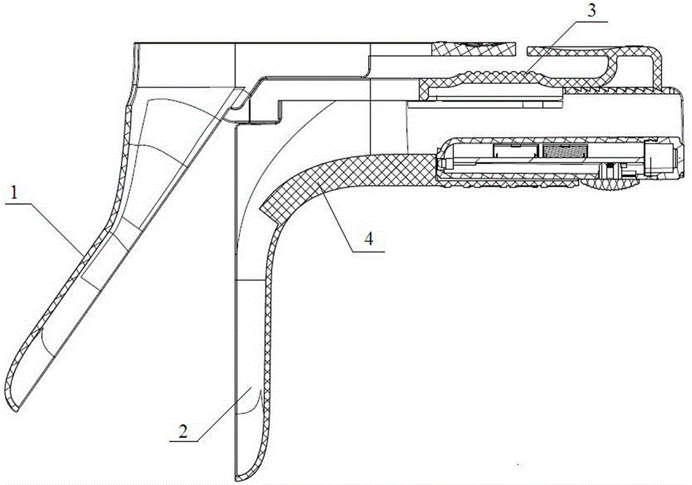
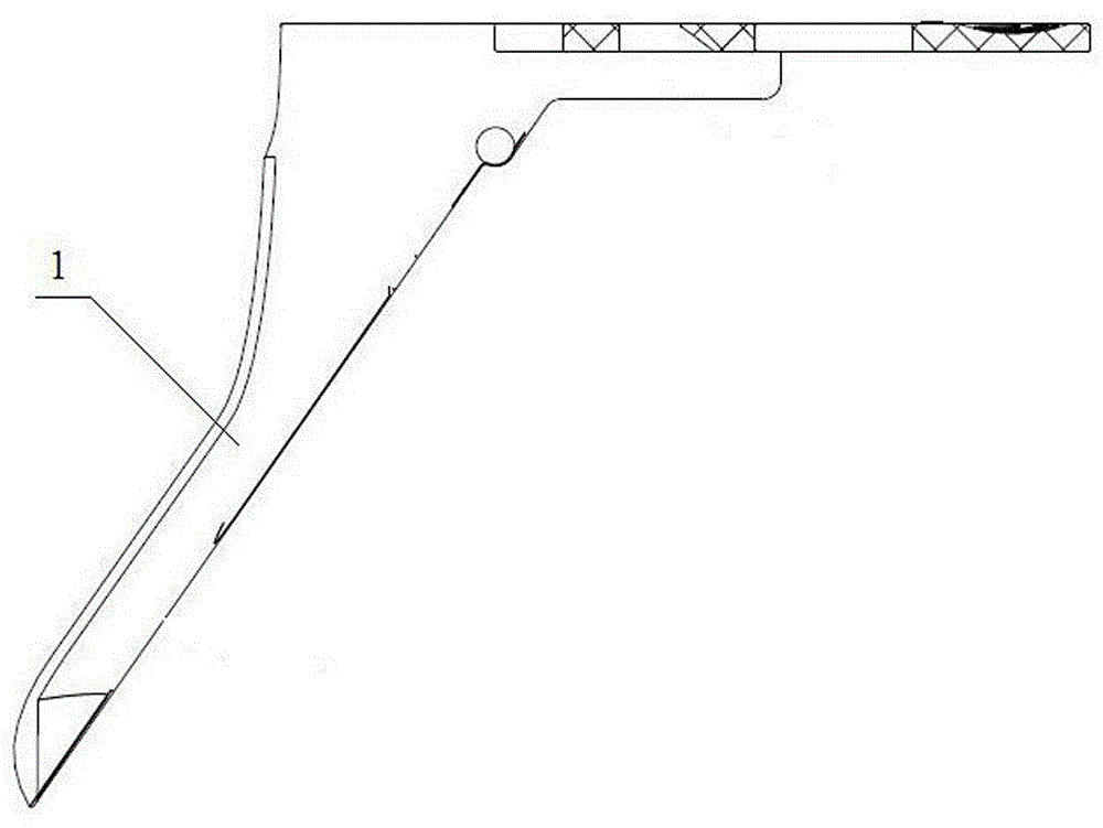
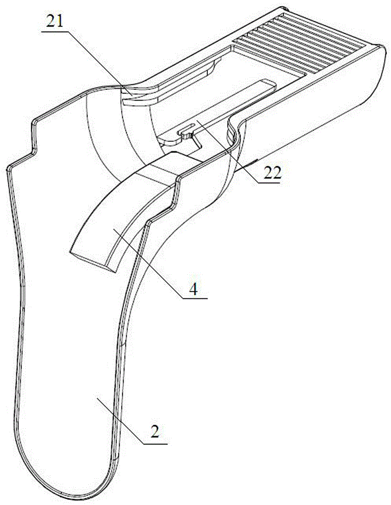
[0028] Such as Figure 1-5 As shown, a gynecological examination system with a biological grade lighting source includes a vaginal expansion mechanism, a lighting mechanism and a diagnostic mechanism. The vaginal expansion mechanism includes an upper expansion leaf 1 and a lower expansion leaf 2, and the upper and lower expansion leaves A handle 3 is also provided. The upper expansion leaf 1 is composed of a duck tongue-shaped expansion part and a manipulation part. Pin holes are respectively provided on both sides of the junction of the expansion part and the manipulation part. The front part of the handle 3 is U-shaped. The two sides of the front end of the handle 3 are correspondingly provided with pin holes, the upper expansion leaf 1 and the handle 3 are hinged through the cooperation of the pin hole and the pin shaft, and the lower expansion leaf 2 is composed of a duck tongue-shaped expansion part and The operating part is composed of an assembly groove, and the lightin...


PUM

PropertyMeasurementUnit
Outer diameteraaaaaaaaaa
The inside diameter ofaaaaaaaaaa
Lengthaaaaaaaaaa

Abstract

The invention relates to a vaginal dilator system with a biological illumination light source. The vaginal dilator system comprises a vaginal dilating mechanism, an illuminating mechanism and a diagnosing mechanism, wherein the vaginal dilating mechanism comprises an upper dilating blade and a lower dilating blade; a handle is also arranged between the upper dilating blade and the lower dilating blade; the upper dilating blade is connected to the handle in a hinging way; the lower dilating blade is connected with the handle in a buckling way; an assembling groove is formed in the tail part of the lower dilating blade; the illuminating mechanism is fixedly arranged in the assembling groove; an arc light-guiding post is also fixedly arranged on the inner side of the lower dilating blade; the end part of the arc light-guiding post is connected with the light-emitting end of the illuminating mechanism in a pushing way; the diagnosing mechanism consists of a cotton ball, a conduit, a diagnostic reagent liquid storing bag and a sealing plug; the cotton ball is fixedly arranged at the end part of the conduit; the conduit and the sealing plug are detachably connected with the diagnostic reagent liquid storing bag; a diagnostic reagent is stored in the diagnostic reagent liquid storing bag. By the vaginal dilator system, the pain of a patient can be greatly relieved, and the vaginal dilator system is good in safety; the vaginal dilator system is convenient to use, and high in diagnosing process efficiency due to the fact that the diagnostic reagent is formulated and bagged in advance.

Description

technical field [0001] The invention relates to a gynecological examination system with a biological grade lighting source, which belongs to the technical field of medical devices. Background technique [0002] At present, disposable vaginal dilators are often used in clinical gynecological examinations. The current vaginal dilator is a very thin duckbill clip. The doctor places it in the patient's vagina and opens the entire clip, and then uses a flashlight or fingers or instruments to cooperate with the inspection. It has the following disadvantages: in gynecological examination or surgery, it is inconvenient to operate, and the light is not easy to reach the affected area during operation. In most cases, it can only be performed blindly. Physicians can only operate with hand feeling or experience, and the diagnosis process takes a long time. It brings great pain to patients, and for diseases that require visual diagnosis, it will lead to inaccurate judgment and easily lea...

Claims


Application Information

Patent Timeline
IPC IPC(8): A61B1/32A61B1/07A61B1/303A61B5/00
Inventor 吕雯岑松原马超武张俊峰刘豪爽
Owner 爱光生物医药(南昌)有限公司
Learn More