Eye fundus examination camera lens

A fundus examination and lens technology, applied in the field of medical devices, can solve the problems of poor component flexibility, inability to complete microscopic imaging, complex structure, etc., and achieve the effect of convenient operation

Active Publication Date: 2013-05-15
SUZHOU KANGJIE MEDICAL
View PDF10 Cites 0 Cited by
  • Summary
  • Abstract
  • Description
  • Claims
  • Application Information

AI Technical Summary

Problems solved by technology

[0002] In the prior art, the lenses used to check the fundus are mostly combined, and are generally used in combination with an optical illuminator, a single aspheric eyepiece lens and a microscopic imaging objective lens. Spherical eyepiece objective lens illuminates and provides light source, microscopic imaging cannot be completed
The above-mentioned combined lens has a complex structure and a large volume. After debugging and assembly, it is an integral type. Users cannot flexibly assemble and debug, and the flexibility between components is poor.

Method used


Image

  • Eye fundus examination camera lens
  • Eye fundus examination camera lens

Examples


Embodiment Construction

[0011] The preferred embodiments of the present invention will be described in detail below in conjunction with the accompanying drawings, so that the advantages and features of the present invention can be more easily understood by those skilled in the art, so as to define the protection scope of the present invention more clearly.

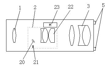
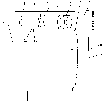
[0012] see figure 1 and figure 2 , the embodiment of the present invention comprises: a kind of fundus inspection lens, comprises: aspheric eyepiece objective lens 1, illumination optical focusing group 2 and microimaging objective lens group 3, is arranged at the rear of aspheric eyepiece objective lens 1 illumination optical focusing Group 2, the microscopic imaging objective lens group 3 is arranged at the rear of the illumination optical focusing group 2, and the illumination optical focusing group 2 includes a reflector 20, a flash lamp, an infrared light emitter 21, a focusing lens group 22 and a focusing motor 23, The reflector 20 is loc...


PUM


Abstract

The invention discloses an eye fundus examination camera lens. The eye fundus examination camera lens comprises an aspheric surface eye contact object lens, a lighting optics focusing set and a microimaging object lens set. The lighting optics focusing set is arranged behind the aspheric surface eye contact object lens. The microimaging object lens set is arranged behind the lighting optics focusing set. The lighting optics focusing set comprises a reflecting lens, a flashlight, an infrared light emitter, a focusing lens set and a focusing motor. The reflecting lens is arranged on the left bottom of the focusing lens set. The flashlight and the infrared light emitter are arranged under the reflecting lens. The focusing motor is arranged above the focusing lens set. The infrared ray emitted by the flashlight and the infrared light emitter penetrates through the reflecting lens and reflected to the aspheric surface eye contact object lens, and passes through the aspheric surface eye contact object lens to project into the eye fundus of an eyeball. The infrared light ray reflected by the eye fundus forms an image through the aspheric surface eye contact object lens. Size of the image can be contracted or expanded in a focusing mode by the focusing lens set. And the image is eventually formed through the microimaging object lens set. With the adoption of the structure, the eye fundus examination camera lens can be conveniently dismounted, flexibly applied, and is strong in replaceability.

Description

technical field [0001] The invention relates to the technical field of medical devices, in particular to a fundus inspection lens. Background technique [0002] In the prior art, the lenses used to check the fundus are mostly combined, and are generally used in combination with an optical illuminator, a single aspheric eyepiece lens and a microscopic imaging objective lens. The spherical eyepiece objective lens illuminates and provides a light source, and microscopic imaging cannot be completed. The above-mentioned combined lens has a complex structure and a large volume. After debugging and assembly, it is an integral type, and the user cannot flexibly assemble and debug, and the flexibility between the components is poor. Contents of the invention [0003] The technical problem mainly solved by the present invention is to provide a fundus inspection lens, which is easy to disassemble, flexible to use, and highly replaceable. [0004] In order to solve the above-mention...

Claims


Application Information

Patent Timeline
IPC IPC(8): A61B3/13A61B3/14
Inventor 朱永林
Owner SUZHOU KANGJIE MEDICAL
Why Eureka
Try Eureka