A visual fundus examination instrument

A fundus inspection and fuselage technology, which is applied in the field of medical devices, can solve problems such as inaccurate diagnosis results, no automatic recording and storage function of the inspection instrument, and easy to cause visual fatigue, etc., and achieve the effect of reducing the number of components equipped

Active Publication Date: 2015-11-11
SUZHOU KANGJIE MEDICAL
View PDF10 Cites 0 Cited by
  • Summary
  • Abstract
  • Description
  • Claims
  • Application Information

AI Technical Summary

Problems solved by technology

[0002] At present, when the traditional fundus examination instrument is in operation, the examinee performs fundus imaging through the fundus examination mirror and optical lighting lamp. The operator's eyes need to be aligned with a pair of microscopes to see the fundus imaging. When imaging, the eyes are always facing the microscope lens. When the operation time is long, it is easy to cause visual fatigue, and the diagnostic results will not be accurate enough. In addition, the diagnostic results are recorded manually, and the inspection instrument itself has no automatic recording or storage function.

Method used


Image

  • A visual fundus examination instrument
  • A visual fundus examination instrument

Examples


Embodiment Construction

[0012] The preferred embodiments of the present invention will be described in detail below in conjunction with the accompanying drawings, so that the advantages and features of the present invention can be more easily understood by those skilled in the art, so as to define the protection scope of the present invention more clearly.

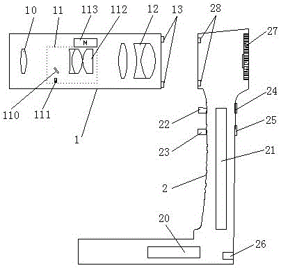
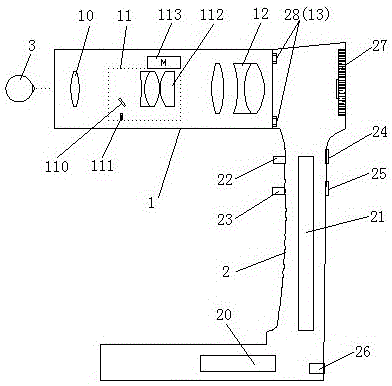
[0013] see figure 1 , the embodiment of the present invention includes: a visual fundus inspection instrument, including: a fundus inspection lens 1 and a body 2, the body 2 is an L-shaped structure, a battery 20 and a control circuit board 21 are arranged inside the body 2, the body 2 is provided with a power switch 22, a shutter button 23, an infrared brightness adjustment button 24 and a focus button 25, a data transmission interface 26 is provided at the rear of the fuselage 2, and a digital camera for infrared reception is provided at the head of the fuselage 2. 27 and the first circuit control line port 28, and the control circuit board 21 ...


PUM


Abstract

The invention discloses a visible fundus examination tester which comprises a fundus examination lens and a tester body. An infrared receiving digital camera and a first circuit control wiring port are arranged on the head portion of the tester body, the fundus examination lens comprises an aspheric surface eye objective lens, a lighting optics focusing set and a micro imaging objective lens set. A second circuit control wiring port is arranged behind the micro imaging objective lens set, and the fundus examination lens is connected with the digital camera through the butting joint of the first circuit control wiring port and the second circuit control wiring port. The infrared rays sent by a flashlight and an infrared light emitter are reflected to the aspheric surface eye objective lens through a reflection mirror, and are projected into the fundus of an eyeball through the aspheric surface eye objective lens. The infrared rays reflected by the fundus are imaged to the digital camera through the imaging of the aspheric surface eye objective lens and the micro imaging objective lens set. Through the mentioned mode, the visible fundus examination tester can catch sight of a fundus imaging picture intuitively, and shoot accurately, and carry out storage or printing.

Description

technical field [0001] The invention relates to the technical field of medical devices, in particular to a visual fundus inspection instrument. Background technique [0002] At present, when the traditional fundus examination instrument is in operation, the examinee performs fundus imaging through the fundus examination mirror and optical lighting lamp. The operator's eyes need to be aligned with a pair of microscopes to see the fundus imaging. When imaging, the eyes are always facing the microscope lens. When the operation time is long, it is easy to cause visual fatigue, and the diagnosis result may not be accurate enough. In addition, the diagnosis result is recorded manually, and the inspection instrument itself has no automatic recording or storage function. Contents of the invention [0003] The main technical problem to be solved by the present invention is to provide a visual fundus inspection instrument, which can intuitively see the imaging pictures of the fundus...

Claims


Application Information

Patent Timeline
Patent Type & Authority Patents(China)
IPC IPC(8): A61B3/13A61B3/14
Inventor 朱永林
Owner SUZHOU KANGJIE MEDICAL
Why Eureka
Try Eureka