
Medical image processing apparatus and ultrasonic imaging apparatus

A processing device, a technology for medical images, applied in image data processing, ultrasound/sonic/infrasound image/data processing, ultrasound/sonic/infrasonic Permian technology, etc. Issues such as phase, time and phase, preparation accuracy, liver tumor accuracy, etc.

Inactive Publication Date: 2010-07-21
TOSHIBA MEDICAL SYST CORP
View PDF1 Cites 13 Cited by
  • Summary
  • Abstract
  • Description
  • Claims
  • Application Information

AI Technical Summary

Problems solved by technology

In this way, the time phase of the boundary between the arterial phase and the portal vein phase cannot be objectively obtained, which may affect the accuracy of creating an arterial phase contrast pattern and the accuracy of determining the type of liver tumor.

Method used


Image

  • Medical image processing apparatus and ultrasonic imaging apparatus
  • Medical image processing apparatus and ultrasonic imaging apparatus
  • Medical image processing apparatus and ultrasonic imaging apparatus

Examples


Embodiment Construction

[0051] A medical image processing device and an ultrasonic image acquisition device according to an embodiment of the present invention will be described. refer to Figure 4 , a medical image processing device according to an embodiment of the present invention will be described. Figure 4 It is a figure which shows the medical image processing apparatus of the Example of this invention.

[0052] The medical image processing apparatus 1 of this embodiment includes an area setting unit 3, an analyzing unit 4, a type judging unit 5, a display control unit 6, a display unit 7, an image storage unit 21, a type contrast mode storage unit 22, and a judgment result storage unit. twenty three. In addition, the medical image processing device 1 is connected to an ultrasonic image acquisition device 8 .

[0053] (ultrasonic imaging device 8)

[0054] The ultrasonic imaging device 8 includes an ultrasonic probe (untrasonic probe). The ultrasonic imaging device 8 transmits ultrasonic...


PUM


Abstract

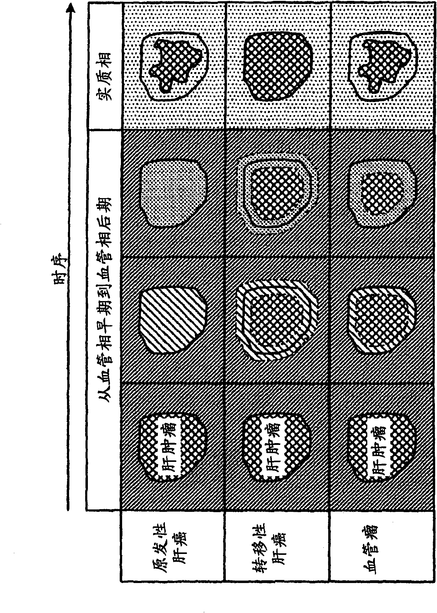
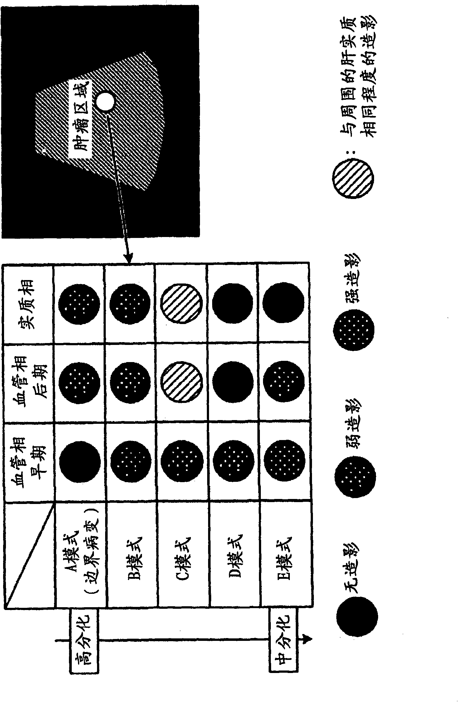
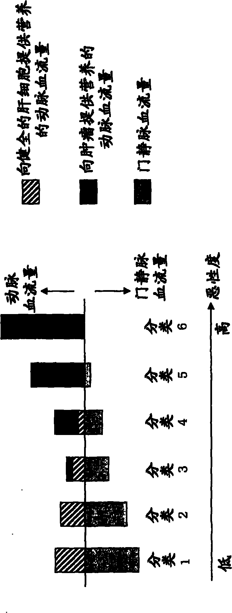
A tumor region setting section sets a liver tumor region for a plurality of ultrasonic image data along a time series acquired by ultrasonically capturing a subject to which a contrast agent has been administered. A TIC generator obtains a time change indicating a time change of the pixel values in the liver tumor region based on the plurality of ultrasonic image data along the time series. A peak-detection section specifies a peak point of the time change and obtains the time and pixel value of that peak point. A first determination section determines the degree of malignancy of the liver tumor based on the time and pixel value of the peak point. A display controller displays the degree of malignancy on a display section.

Description

technical field [0001] The present invention relates to a medical image processing device for judging tumors based on ultrasonic images obtained by photographing a subject to which a contrast agent has been administered. In addition, the present invention also relates to an ultrasonic image acquisition device for judging a tumor based on an ultrasonic image. Background technique [0002] "Malignant neoplasm (cancer)", "heart disease" and "cerebrovascular disease (cerebral stroke)" are among the top three causes of death in Japan . In particular, cancer has the largest number of deaths among the three major causes of death, accounting for 30% of the total number of deaths. [0003] When cancer is classified according to the site of occurrence, in Japan, it can be classified into lung cancer, gastric cancer, colon cancer, and liver cancer in descending order of the number of patients . However, the 5-year relative survival rate (relative survival) of patients with gastric ...

Claims


Application Information

Patent Timeline
Patent Type & Authority Applications(China)
IPC IPC(8): A61B8/08
CPCG06T7/0016G06T2207/10132G06T2207/30056
Inventor 佐藤俊介山形仁
Owner TOSHIBA MEDICAL SYST CORP
Learn More