Intravaginal ultrasonic probe for gynecological operation and examination

A technology for gynecological surgery and ultrasonic probe, applied in the field of medical devices, can solve the problems of inability to adapt to various uterine positions and various gynecological operations, the large size of the B-type ultrasonic monitor probe, the limitations of the B-type ultrasonic monitor, and the like

Inactive Publication Date: 2011-07-20
WUXI CANSONIC MEDICAL SCI & TECH
View PDF8 Cites 0 Cited by
  • Summary
  • Abstract
  • Description
  • Claims
  • Application Information

AI Technical Summary

Problems solved by technology

[0014] There is a demand for implantable B-mode ultrasonic monitors for women who are adapted to the above-mentioned various gynecological operations, but the B-mode ultrasonic monitors of the prior art are still limited.
Or the volume of the B-type ultrasonic monitor probe is large, or it needs a special holder or fixer to fix it, or the position of the B-type ultrasonic monitor probe is difficult to reach a more ideal place, or the B-type ultrasonic monitor The probe cannot scan the complete uterus at a fixed position, so it cannot adapt to various uterine positions and various gynecological operations

Method used


Image

  • Intravaginal ultrasonic probe for gynecological operation and examination
  • Intravaginal ultrasonic probe for gynecological operation and examination
  • Intravaginal ultrasonic probe for gynecological operation and examination

Examples


Embodiment Construction

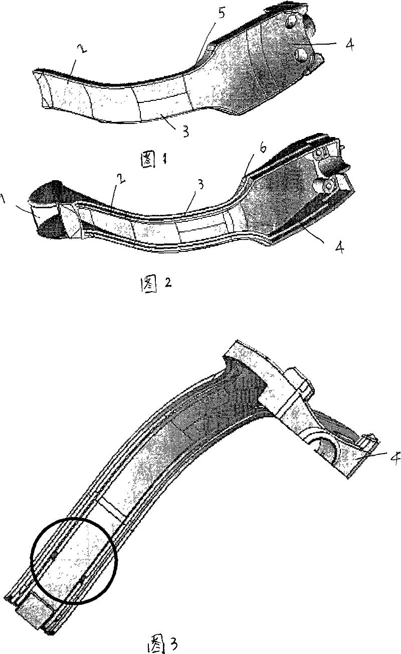
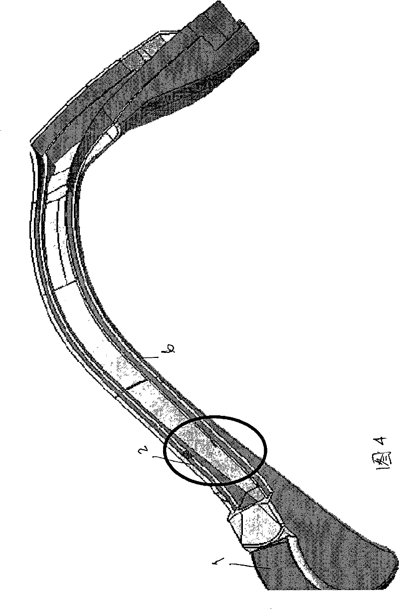
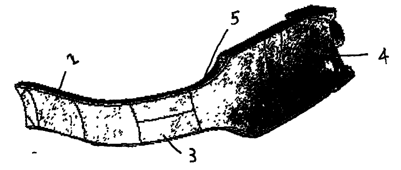
[0027] Embodiments are described below in conjunction with the accompanying drawings. Wherein the device of each embodiment can make it easy for the surgeon to align the medical equipment used with the ultrasonic transmitter, the B-ultrasound transducer 1, the probe arm 2 covering the flexible lead-out wire, the cable lead-out arm 5 and the arc connection rod housing). The probe arm and the cable lead-out arm pass through an arc-shaped connecting rod, considering that the length of the probe arm is

[0028] According to this embodiment, when inserting the medical device through the patient's cervix, the surgeon can ensure that the medical device is positioned parallel to the extension, thereby "putting" or "putting" the medical device into the ultrasound beam.

[0029] The other end of the present invention is also provided with a cable connector, which is connected with the host through a cable.

[0030] The cross-section of the probe arm 2 or the arc connecting rod 3 is pr...


PUM


Abstract

The invention discloses an intravaginal ultrasonic probe for gynecological operation and examination, comprising a B ultrasonic transducer arranged in the vagina of a patient during in-utero, uterine neck and uterine tube surgery operations, a probe arm for connecting the B ultrasonic transducer and a cable outgoing arm, wherein the probe arm and the cable outgoing arm are transited through an arc connection rod, the probe is installed at the top end of the probe arm, and the extension lines of the probe arm and the cable outgoing arm form an included angle of 78-87 degrees. The probe arm andthe arc connection rod and even the cable outgoing arm are all flat and wide cable sheath coated with flexible circuit boards, the sections of the probe arm and the arc connection rod are matched with the lower piece of a speculum, and the cable outgoing arm is connected with a host through a connection cable.

Description

technical field [0001] The invention relates to a medical instrument for gynecological operation and gynecological examination, especially an intravaginal real-time ultrasonic detection device including uterine, cervix and fallopian tube auxiliary operations. Background technique [0002] Gynecological examinations and intrauterine, cervical, and fallopian tube surgical procedures have traditionally relied solely on the "feel" transmitted through manually operated instruments and the experience of the surgeon. However, if the doctor is not sure about the location and size of the uterus, it may puncture the uterus or cause other harm to the body, such as in the case of cervical stenosis or uterine tumors (endometrioma or sarcoma). more dangerous. The main risks of uterine penetration mentioned above include bleeding, trauma to abdominal viscera, and damage to internal organs such as the bowel, omentum, mesentery, ureters, and fallopian tubes. Therefore, laparoscopy or lapar...

Claims


Application Information

Patent Timeline
Patent Type & Authority Patents(China)
IPC IPC(8): A61B8/12
CPCA61B8/12A61B17/42A61B8/445
Inventor 王敏岐
Owner WUXI CANSONIC MEDICAL SCI & TECH
Why Eureka
Try Eureka