Quantification and display of cardiac chamber wall thickening

A technique of wall thickness, myocardium, in the field of medical diagnostic ultrasound systems

Active Publication Date: 2009-06-10
KONINKLIJKE PHILIPS ELECTRONICS NV
View PDF6 Cites 14 Cited by
  • Summary
  • Abstract
  • Description
  • Claims
  • Application Information

AI Technical Summary

Problems solved by technology

Moreover, the Doppler method can

Method used


Image

  • Quantification and display of cardiac chamber wall thickening
  • Quantification and display of cardiac chamber wall thickening
  • Quantification and display of cardiac chamber wall thickening

Examples


Embodiment Construction

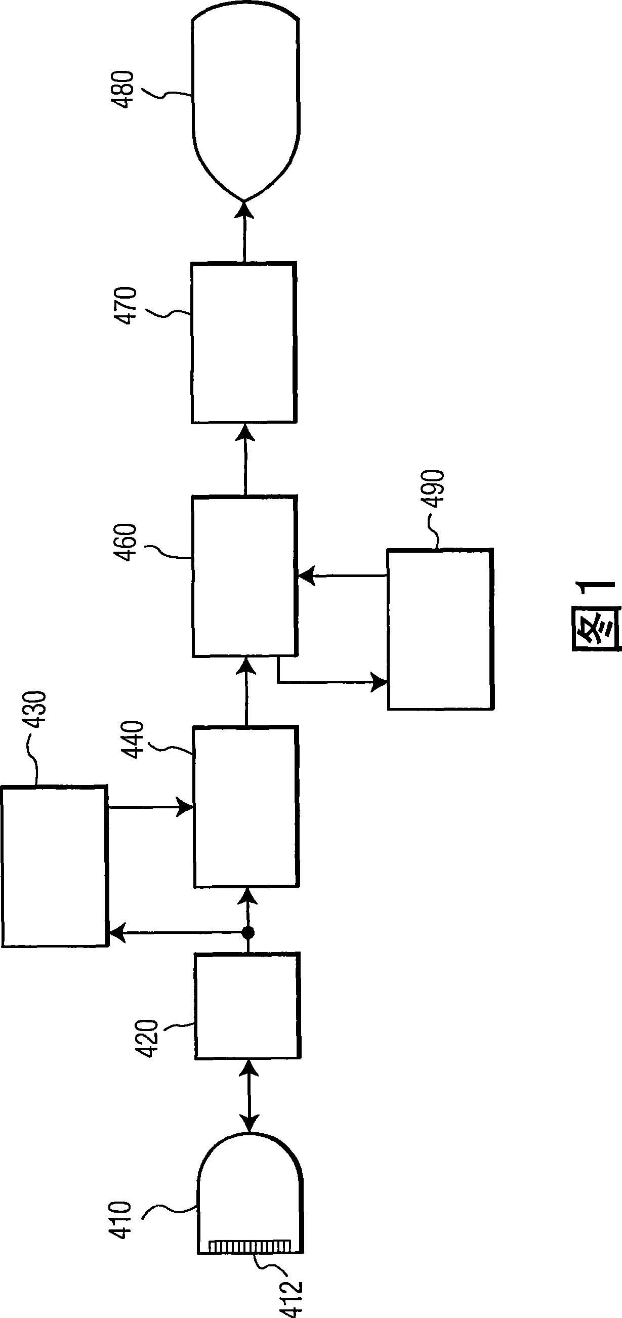
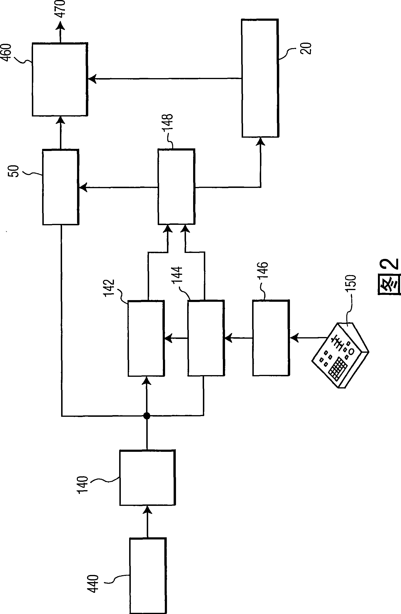
[0014] First, reference is made to FIG. 1 , which shows in block diagram form an ultrasonic diagnostic imaging system constructed in accordance with the principles of the present invention. A probe or scan head 410 comprising a one-dimensional (1D) or two-dimensional (2D) array 412 of transducer elements transmits ultrasound waves and receives ultrasound echo signals. This transmission and reception is performed under the control of the beamformer 420, which processes echo signals received from the body being scanned to form coherent beams from the echo signals. When it is desired to present Doppler information, the echo information is Doppler processed by the Doppler processor 430, and the processed Doppler information is coupled to form a 2D or 3D Doppler image image processor 440 . For B-mode imaging of tissue structures, the echo signal is image processed by amplitude detection and scan converted to the desired image format for display. The image is passed through memo...


PUM


Abstract

An ultrasonic imaging system and method are described for quantification and display of myocardial wall thickening. The endocardial and epicardial borders in an image sequence are defined over a heart cycle and changes in the distance between the borders are tracked at specified locations around the myocardium over the heart cycle, The changes in distance are presented to the user in a graphical format, preferably together with another measure of the cardiac cycle such as chamber volume variation, ejection fraction, or the ECG waveform. The changes in the distance of chord lengths across the myocardium provide a direct indication of wall thickness variation at the specified locations. Preferably the tracking of the specified locations over the heart cycle is done by speckle tracking. The inventive technique can also represent strain at the specified locations of the myocardium.

Description

[0001] This application claims the benefit of US Provisional Patent Application Nos. 60 / 803148 and 60 / 823114, filed May 25, 2006, and August 22, 2006, respectively. technical field [0002] The present invention relates to medical diagnostic ultrasound systems, and more particularly, to ultrasound systems for performing quantitative echocardiographic measurements. Background technique [0003] There are many methods for quantifying cardiac chamber function. Ventricular wall motion indicative of myocardial infarction and ejection fraction is an essential diagnostic tool of the echocardiographer. These diagnostic tools require that the ventricles be delineated and tracked over a series of images over the cardiac cycle to complete measurements of the delineated myocardium and ventricles. Techniques for delineating the heart wall in ultrasound images include, for example, automatic border tracking of the blood-tissue interface and tissue Doppler imaging of heart wall motion for...

Claims


Application Information

Patent Timeline
IPC IPC(8): G01S7/52
CPCA61B8/0858G06T7/20G06T7/0012G06T2207/30048G01S7/52073G01S7/52071G01S7/52087G01S7/52042A61B8/00G01S15/00
Inventor I·萨尔戈S·塞特尔迈尔
Owner KONINKLIJKE PHILIPS ELECTRONICS NV
Why Eureka
Try Eureka