


Quick Research
Generate reliable direction feasibility study reports for your R&D in just a few steps.

Technical Q&A
Discover and master advanced knowledge NOW. Basics, ideas, possibilities, all at once.

Find Solutions
As an expert in R&D theories, this can generate solutions to your technical problems instantly.

Evaluate Feasibility
Analyze your overall solution with one click, know your potential R&D risks in advance.

Monitor Landscape
Get weekly tech updates, stay abreast of the latest tech innovations and key insights.
Method and kit to profile tumours by biomarker analyses including transcriptional factor assays
A technology of transcription factors and biomarkers, applied in the fields of biochemical equipment and methods, analytical materials, anti-tumor drugs, etc., can solve the problem of not providing tumor profiles and other problems
- Summary
- Abstract
- Description
- Claims
- Application Information
AI Technical Summary
Problems solved by technology
Method used
Image

Examples
Embodiment 1
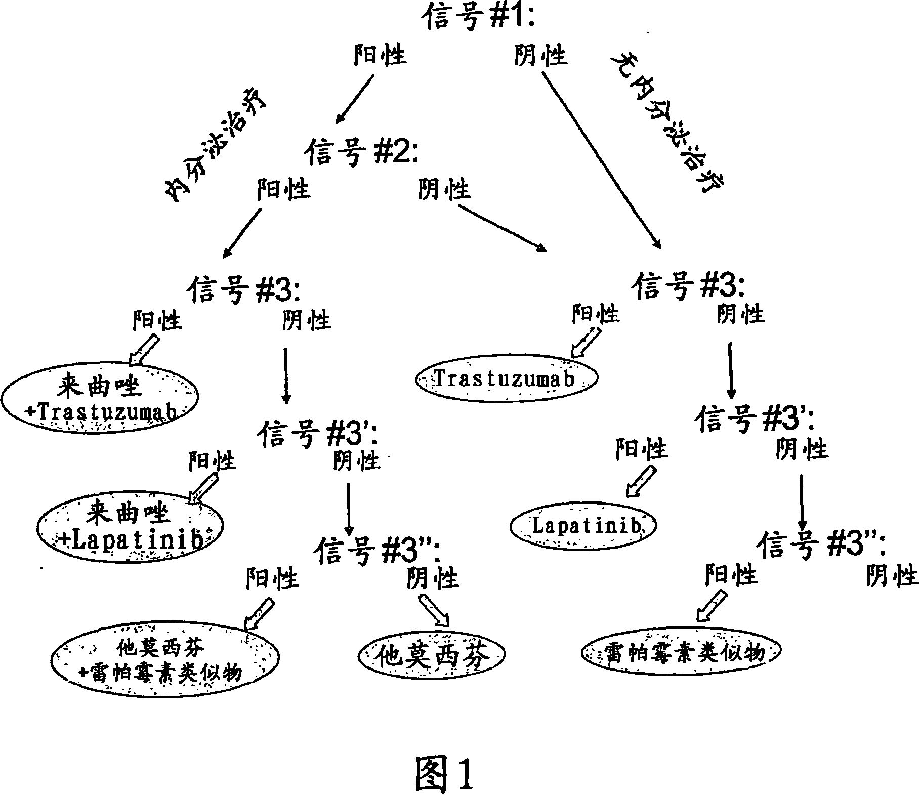
2. Example 1: Method for Classifying Breast Tumor Responsiveness to Different Drugs
[0133] Two microarrays were spotted on glass slides: the first microarray consisted of triplicate spots of double-stranded DNA capture probes containing binding sites specific for ERα. The sequence of the sense strand is as follows: 5'-CAGGTCACAGTGACCTGATCAAAGTT-3'. The second microarray consisted of triplicate spots of protein capture probes corresponding to antibodies specific for Erα, ErbB1, ErbB2 and mTOR, respectively. A hybridization chamber was attached to a glass slide to physically separate the two microarrays.
[0134] The first microarray was contacted with protein extracts of breast tumor samples diluted in binding buffer consisting of Hepes buffer supplemented with salt, EDTA, glycerol, protease and phosphatase inhibitors. A second microarray was exposed to protein extracts of the same breast tumor samples diluted in MOPSO buffer supplemented with salts, detergents and proteas...
Embodiment 2
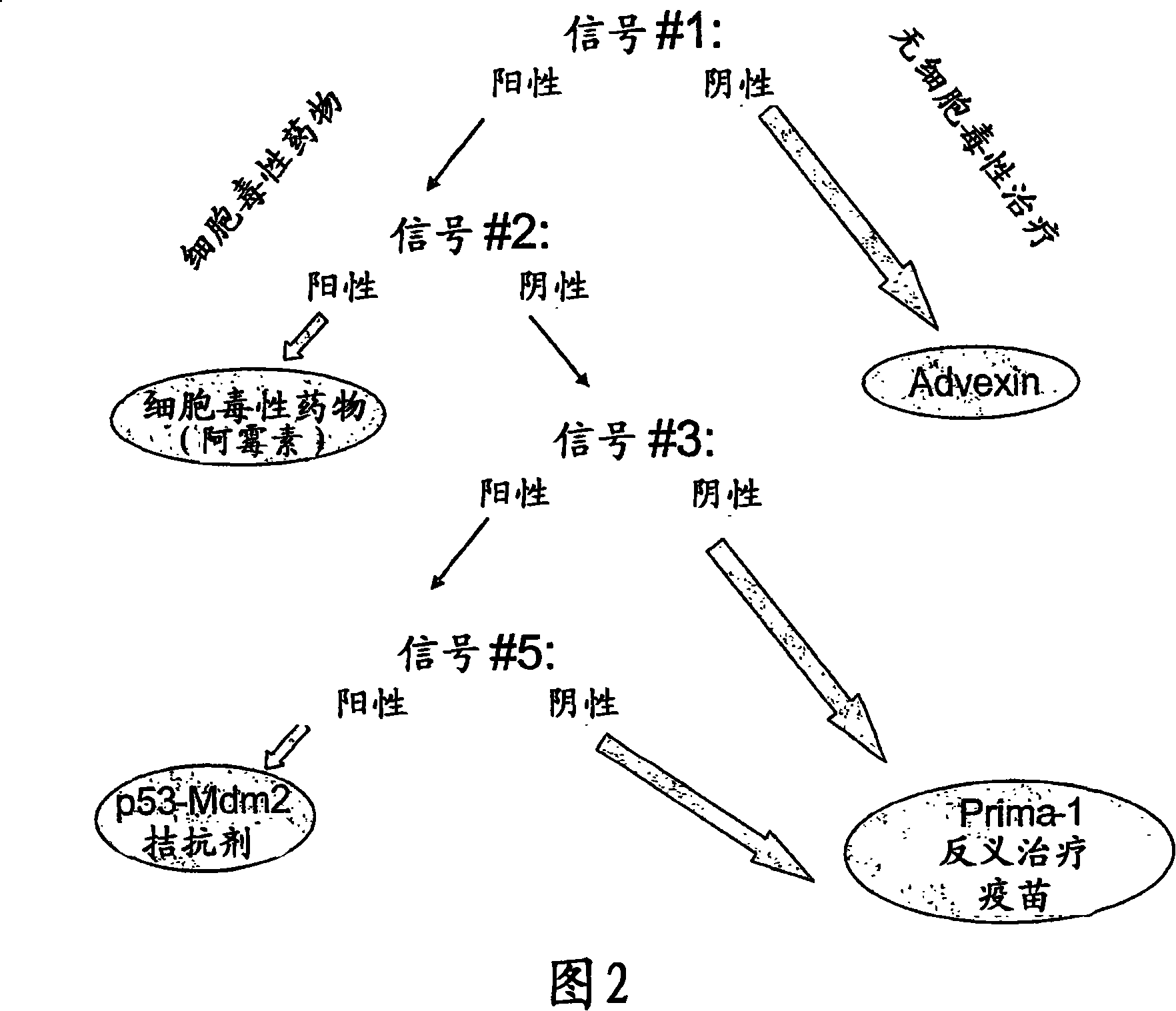
3. Example 2: Method of Analyzing Tumors for Tailored p53 Therapy
[0141] Each well of the 96-well plate was coated with different capture molecules: the first group of 5 wells was coated with p53 monoclonal antibody; the second group of 5 wells coated with streptavidin was coated with p53-specific Binding sites are coated with biotinylated dsDNA capture probes. The sequence of the sense strand is as follows: 5'-CTTGGACATGCCCGGGCATGTCCCTC-3'; the 5 wells of the third group were coated with the monoclonal antibody of Mdm2; the 5 wells of the fourth group were coated with the p53 as used in the first group Antibody coating.
[0142] Two wells of each set were exposed to binding buffer only ("blank") and the remaining 3 wells of each set were exposed to protein extracts of test samples diluted in binding buffer ("test"). Binding buffer for groups 1, 3 and 4 consisted of MOPSO buffer containing salts, detergents, proteins and protease inhibitors. The binding buffer used for g...
Embodiment 3
4. Example 3: Other tumor profiling
[0150] The inventors have also obtained other tumor profiling based on the method according to the invention, which allows the treatment of specific cancers.
a) Angiogenesis:
[0151] The growth of new vessels from preexisting vessels is considered an important event essential for tumor survival and growth. Anti-angiogenic therapies have thus been developed that target angiogenic tumor biomarkers such as the transcription factor HIF1α. The presence and DNA binding activity of HIF1α (monitored by signals #1 and #2 of the present invention) would indicate the use of anti-angiogenic molecules, such as the specific HIF1α inhibitor astatin. However, binding of p53 to HIF1α results in the inactivation of HIF1α. Thus monitoring the presence or absence of p53 via signal #3 and the p53-HIF1α interaction via signal #5 may not help give any precise information about the anti-angiogen (ie, the molecule used), but will enable the clinician Abando...
PUM
 Abstract
Description
Claims
Application Information
 - R&D Engineer
- R&D Manager
- IP Professional
- Industry Leading Data Capabilities
- Powerful AI technology
- Patent DNA Extraction
Browse by: Latest US Patents, China's latest patents, Technical Efficacy Thesaurus, Application Domain, Technology Topic, Popular Technical Reports.
© 2024 PatSnap. All rights reserved.Legal|Privacy policy|Modern Slavery Act Transparency Statement|Sitemap|About US| Contact US: help@patsnap.com