
Method and apparatus for detecting acceleration field of tissue image of colorful Doppler ultrasonography

A technology of Doppler velocity and Doppler map is applied in the field of acceleration field detection of tissue Doppler map in color Doppler, and can solve the problems such as inability to obtain

Inactive Publication Date: 2008-09-17
FUZHOU UNIV
View PDF3 Cites 0 Cited by
  • Summary
  • Abstract
  • Description
  • Claims
  • Application Information

AI Technical Summary

Problems solved by technology

However, in the prior art, the acceleration field of the tissue Doppler velocity field, that is, the acceleration detection at each moment cannot be obtained.

Method used


Image

  • Method and apparatus for detecting acceleration field of tissue image of colorful Doppler ultrasonography
  • Method and apparatus for detecting acceleration field of tissue image of colorful Doppler ultrasonography
  • Method and apparatus for detecting acceleration field of tissue image of colorful Doppler ultrasonography

Examples


Embodiment Construction

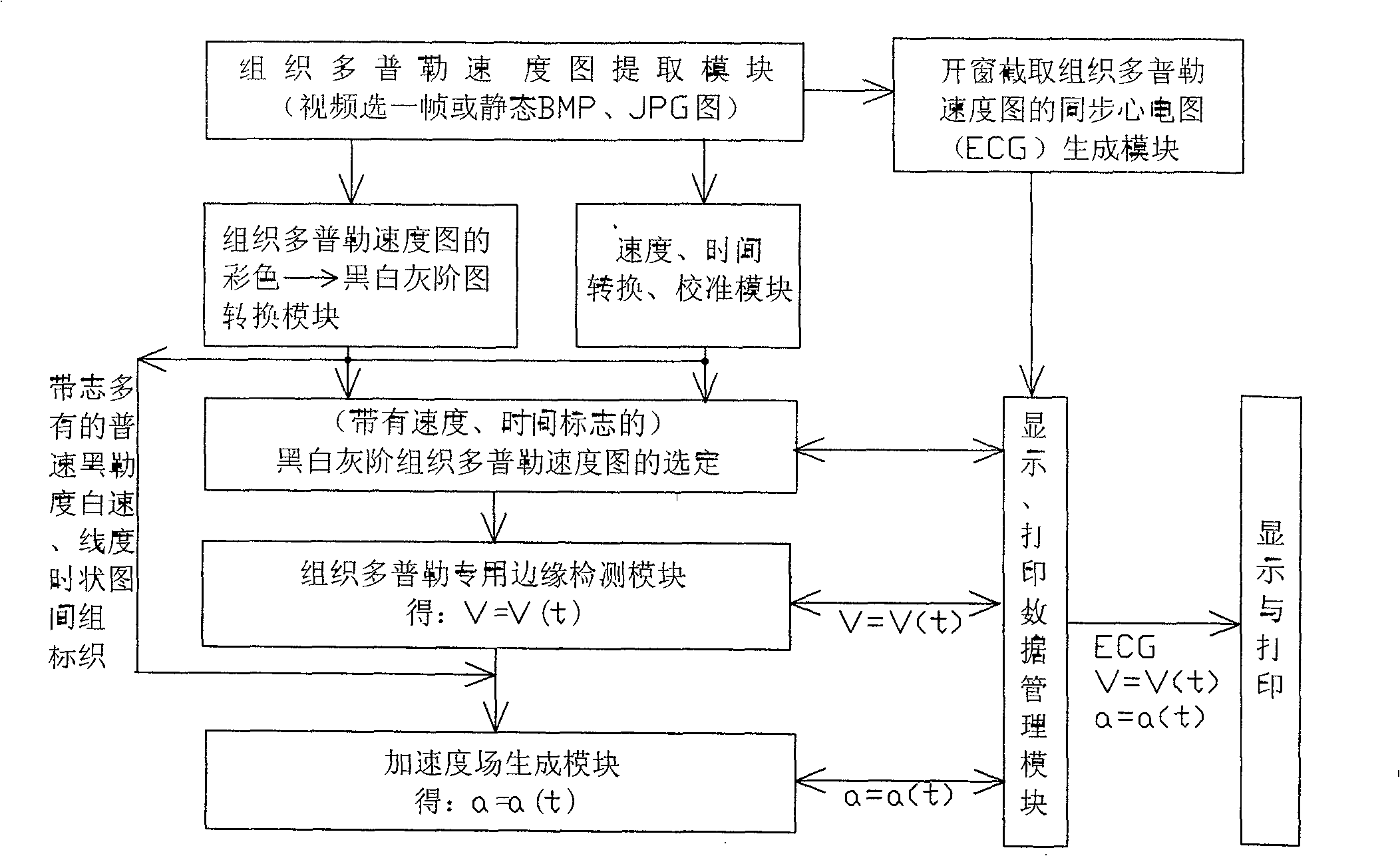
[0021] like figure 1 As shown in -5, the acceleration field detection method of the tissue Doppler image in the color ultrasound of the present invention is as follows:

[0022] It includes any one or more of the following four methods, each with the following steps:

[0023] (1) The first method

[0024] (a) The tissue Doppler color velocity map is extracted by the tissue Doppler velocity map extraction module,

[0025] (b) Converting into a black and white image through the color→black and white grayscale image conversion module of the tissue Doppler velocity map in step (a),

[0026] (c) another routing step (a) through the speed, time conversion, calibration module,

[0027] (d) through steps (b) and (c), the selection of the black-and-white gray-scale tissue Doppler velocity map with velocity and time markers is performed together,

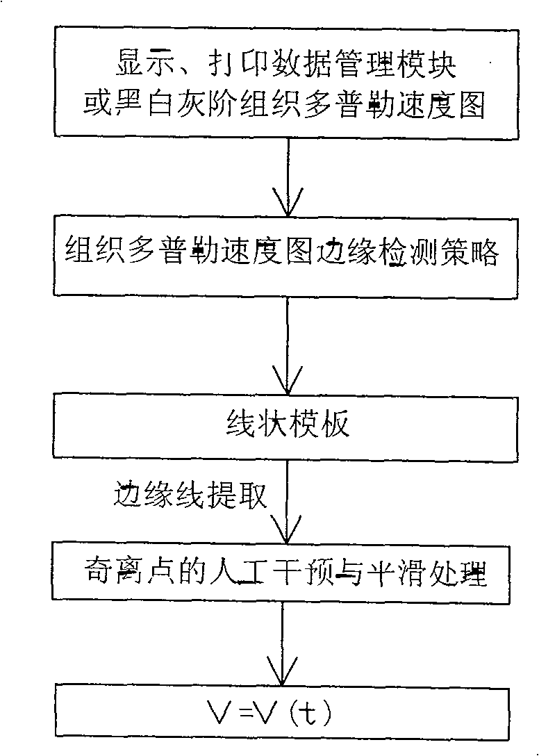
[0028] (e) After the detection of the special edge detection module of tissue Doppler, V=V(t) is obtained,

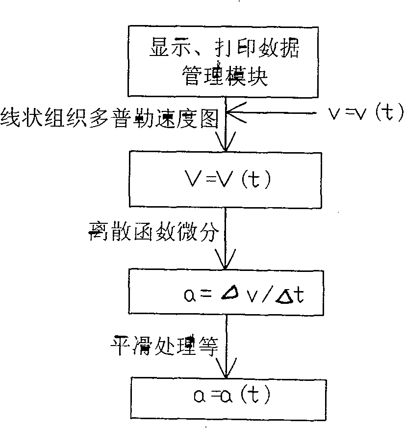
[0029] (f) After the accel...


PUM


Abstract

The invention relates to a detecting method of tissue Doppler acceleration field and its device in color super technique, it is about abstracting rate curve V=V(t) in some position of the heart constructions from tissue Doppler velocity diagram, doing first-order differentiation to it and getting the detection of acceleration field of the acceleration a=a(t) in said position of the heart conditions. Said invention and its device could detect the acceleration field while moving in said potion at the base of getting the velocity diagram of heart constructions in some position in color super new tech tissue Doppler detection. According to Newton's second law F=ma, the detection of acceleration offers very important information to the forced situation in said position, therefore, it has very important values to homodynamic research in different positions of the heart construction and the diagnosis of cardiovascular diseases.

Description

Technical field: [0001] The invention relates to an acceleration field detection method and a device, in particular to an acceleration field detection method and a device for tissue Doppler images in color ultrasound. Background technique: [0002] A relatively new technology in imported color ultrasound is tissue Doppler map, which is the tissue velocity field map of the heart structure. , we will refer to it as the velocity field for short, and the speed of the national invention patent "Detection method and device for velocity field and acceleration field of omnidirectional M-mode cardiogram" (publication number: CN1539379A; application number: 200310104284.0) Field has a similar meaning. However, in the prior art, the acceleration field of the tissue Doppler velocity field, that is, the acceleration detection at every moment, cannot be obtained. [0003] It can be known from Newton's second law F=ma. The detection of the acceleration field will bring people a big step...

Claims


Application Information

Patent Timeline
Patent Type & Authority Patents(China)
IPC IPC(8): A61B8/00A61B5/00G01P15/00
Inventor 林强
Owner FUZHOU UNIV
Learn More