
Registration maps using intra-cardiac signals

a technology of intra-cardiac signals and register maps, which is applied in the field of medical imaging systems, can solve the problems that the register of electroanatomical maps of the heart with anatomic landmarks may not always be optimal, and achieve the effect of improving the accuracy of the register

Active Publication Date: 2018-05-01
BIOSENSE WEBSTER (ISRAEL) LTD
View PDF26 Cites 36 Cited by
  • Summary
  • Abstract
  • Description
  • Claims
  • Application Information

AI Technical Summary

Benefits of technology

The method improves the accuracy of catheter placement and navigation by aligning electroanatomic maps, enabling precise identification of points of interest and facilitating effective treatment of cardiac arrhythmias by ensuring compatible unit measurements and interoperative maps.

Problems solved by technology

Registration of electroanatomical maps of the heart with anatomic landmarks may not always be optimum.

Method used


Image

  • Registration maps using intra-cardiac signals
  • Registration maps using intra-cardiac signals
  • Registration maps using intra-cardiac signals

Examples


example 1

[0061]Reference is now made to FIG. 6, which shows two electroanatomic maps 67, 69 of the heart, which are to be placed in registration, in accordance with an embodiment of the invention. As the maps 67, 69 were prepared by different systems, their coordinate systems and scales are generally not identical. It will be noted by reference to the intervals 71, 73 that the scales of the maps 67, 69 differ. Moreover, the axes of rotation of the maps 67, 69 are not identical, as shown by intersecting lines 75, 77. Map 67 illustrates local activation times, and was prepared using a phase analysis mode of the above-noted CARTO system. Map 69 is an isochronous map that was prepared using a phase analysis mode of the AMYCARD-01C diagnostic system.

[0062]Points used to place the maps into registration can be appreciated on both maps 67, 69. For example, points 79, 81, 83 on map 67 and points 85, 87, 89 on map 69 all show corresponding electrical events that correspond anatomically to myocardium ...


PUM


Abstract

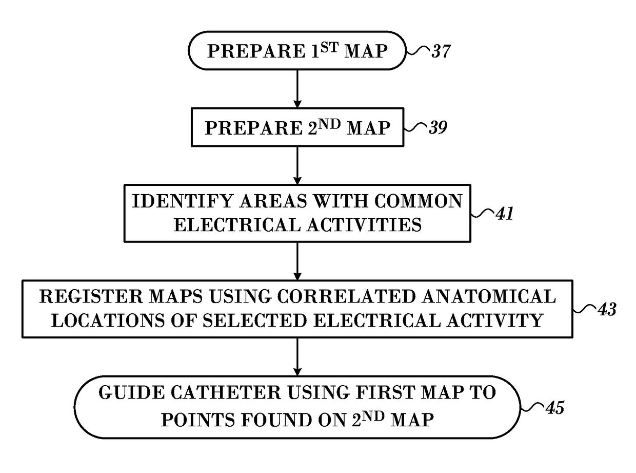
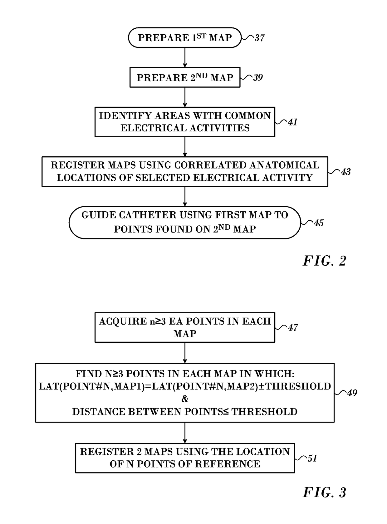
Cardiac catheterization is facilitated by generating first and second electroanatomic maps of a heart of a subject and designating common spatial locations that correspond to first electrical events on the first electroanatomic map and second electrical events on the second electroanatomic map. The common spatial locations of the first electroanatomic map and the second electroanatomic map are aligned to establish an aligned map, and using the location data on the aligned map to guide a probe to a point of interest.

Description

BACKGROUND OF THE INVENTION[0001]Field of the Invention[0002]This invention relates to medical imaging systems. More particularly, this invention relates to improvements in medical image analysis.[0003]Description of the Related Art[0004]Three-dimensional (3-D) images of internal organs are useful in many catheter-based diagnostic and therapeutic applications, and real-time imaging is widely used during surgical procedures. Ultrasound imaging is a relatively convenient mode of real-time imaging, though the resolution of real-time ultrasound images is generally not as good as the resolution obtained from other imaging modalities, such as computerized tomography (CT) and magnetic resonance 20 imaging (MRI).[0005]Methods for 3-D mapping of a heart using a position-sensing catheter are well known in the art. For example, U.S. Pat. No. 5,738,096 to Ben-Haim, whose disclosure is incorporated herein by reference, describes a position-sensing probe brought into contact with multiple points ...

Claims


Application Information

Patent Timeline
Patent Type & Authority Patents(United States)
IPC IPC(8): A61B5/044A61B5/00A61B5/0452G06F19/00A61B5/042A61B5/06A61B5/04A61B34/20A61B5/046A61B5/361G16B45/00
CPCG16H50/50A61B5/044A61B5/04012A61B5/0422A61B5/0452A61B5/066A61B5/7475A61B5/7485A61B5/068A61B2034/2051A61B5/046A61B5/6852A61B5/7264A61B5/743G16Z99/00A61B5/287A61B5/349A61B5/361A61B5/339A61B5/316
Inventor URMAN, ROYKRUPNIK, RONENMIZRAHI, LIRON SHMUEL
Owner BIOSENSE WEBSTER (ISRAEL) LTD
Learn More