Method and system for automatic detection of spinal bone lesions in 3D medical image data

a 3d computed tomography and bone lesions technology, applied in image analysis, image enhancement, instruments, etc., can solve the problems of manual identifying and volumetric measurement, spinal cord compression with severe neurological impairment, and manual annotation of spinal bone lesions from 3d computed tomography data,

Active Publication Date: 2014-04-08
SIEMENS HEALTHCARE GMBH
View PDF18 Cites 0 Cited by
  • Summary
  • Abstract
  • Description
  • Claims
  • Application Information

AI Technical Summary

Problems solved by technology

Spinal bone lesions may cause debilitating pain, pathologic fractures, and spinal cord compression with sever neurological impairment.
However, reading an manually identifying and volumetrically measuring, i.e. annotating, spinal bone lesions from 3D computed tomography (CT) data is a challenging and labor intensive task, even for expert radiologists.
Further, there may be significant inter- and intra-user variability among manual bone lesion annotations.

Method used


Image

  • Method and system for automatic detection of spinal bone lesions in 3D medical image data
  • Method and system for automatic detection of spinal bone lesions in 3D medical image data
  • Method and system for automatic detection of spinal bone lesions in 3D medical image data

Examples


Embodiment Construction

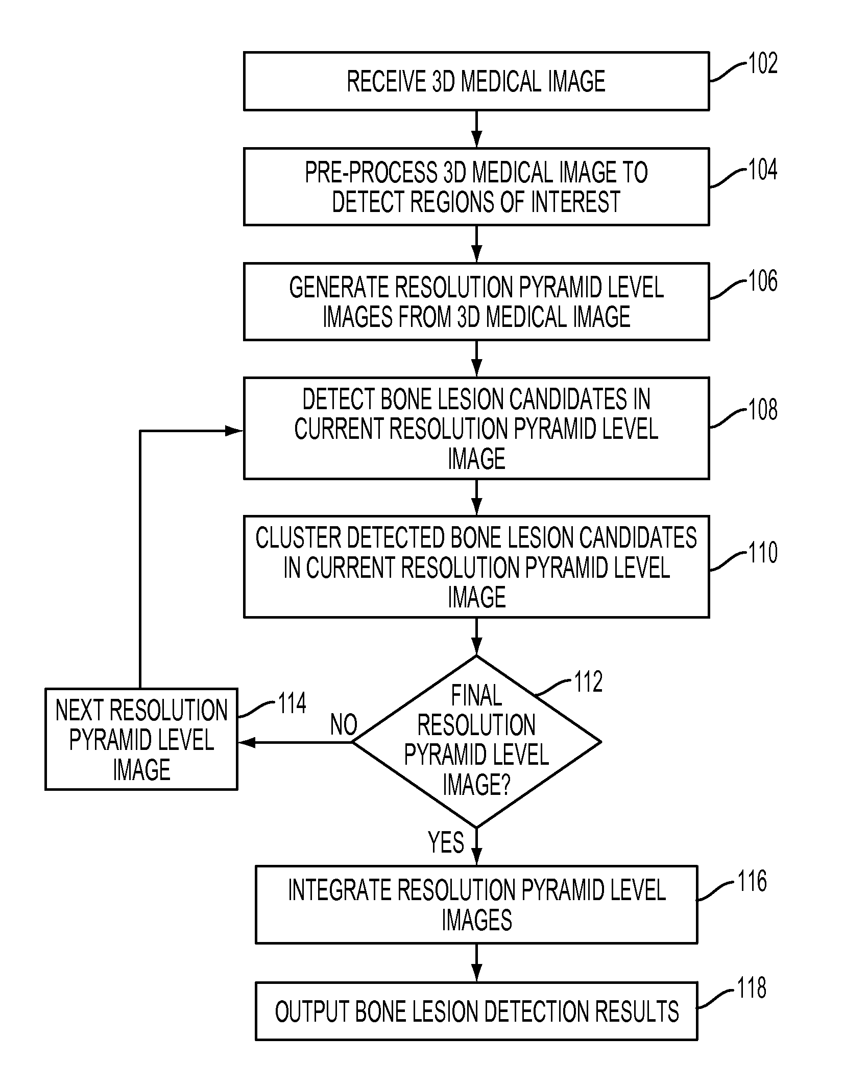
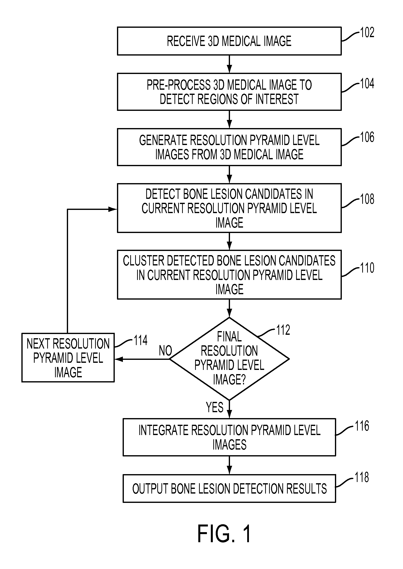
[0012]The present invention is directed to a method and system for automatic detection and volumetric quantification of bone lesions in medical images, such as computed tomography (CT), magnetic resonance (MR) images, etc. Embodiments of the present invention are described herein to give a visual understanding of the bone lesion detection method. A digital image is often composed of digital representations of one or more objects (or shapes). The digital representation of an object is often described herein in terms of identifying and manipulating the objects. Such manipulations are virtual manipulations accomplished in the memory or other circuitry / hardware of a computer system. Accordingly, it is to be understood that embodiments of the present invention may be performed within a computer system using data stored within the computer system.

[0013]Embodiments of the present invention provide fully automatic detection and volumetric measurements of bone lesions in 3D medical image dat...


PUM


Abstract

A method and system for automatic detection and volumetric quantification of bone lesions in 3D medical images, such as 3D computed tomography (CT) volumes, is disclosed. Regions of interest corresponding to bone regions are detected in a 3D medical image. Bone lesions are detected in the regions of interest using a cascade of trained detectors. The cascade of trained detectors automatically detects lesion centers and then estimates lesion size in all three spatial axes. A hierarchical multi-scale approach is used to detect bone lesions using a cascade of detectors on multiple levels of a resolution pyramid of the 3D medical image.

Description

[0001]This application claims the benefit of U.S. Provisional Application No. 61 / 432,612, filed Jan. 14, 2011, the disclosure of which is herein incorporated by reference.BACKGROUND OF THE INVENTION[0002]The present invention relates to detection of bone lesions in medical images, and more particularly, to automatic detection and volumetric quantification of spinal bone lesions in 3D medical images.[0003]Detection and volumetric quantification of spinal bone lesions is important in the treatment of metastizing cancer. Spinal bone lesions may cause debilitating pain, pathologic fractures, and spinal cord compression with sever neurological impairment. In addition to assessing risks of certain courses of the disease, detection and volumetric quantification of bone lesions is important for accurate quantification of disease progression or response to therapy. However, reading an manually identifying and volumetrically measuring, i.e. annotating, spinal bone lesions from 3D computed tom...

Claims


Application Information

Patent Timeline
Patent Type & Authority Patents(United States)
IPC IPC(8): G06K9/70G06K9/46G06K9/68G06K9/00G06K9/62G06V10/774
CPCG06K2209/40G06T2207/10072G06T2207/30012G06K2209/055G06T2207/20081G06K9/4614G06T2207/20016G06T2207/30096G06K9/6256G06T7/0012G06V10/446G06V2201/033G06V2201/12G06V10/774G06F18/214
Inventor WELS, MICHAELSUEHLING, MICHAELZHOU, SHAOHUA KEVINLIU, DAVIDWU, DIJIAALVINO, CHRISTOPHER V.KELM, MICHAELSOZA, GRZEGORZCOMANICIU, DORIN
Owner SIEMENS HEALTHCARE GMBH
Why Eureka
Try Eureka