


Quick Research
Generate reliable direction feasibility study reports for your R&D in just a few steps.

Technical Q&A
Discover and master advanced knowledge NOW. Basics, ideas, possibilities, all at once.

Find Solutions
As an expert in R&D theories, this can generate solutions to your technical problems instantly.

Evaluate Feasibility
Analyze your overall solution with one click, know your potential R&D risks in advance.

Monitor Landscape
Get weekly tech updates, stay abreast of the latest tech innovations and key insights.
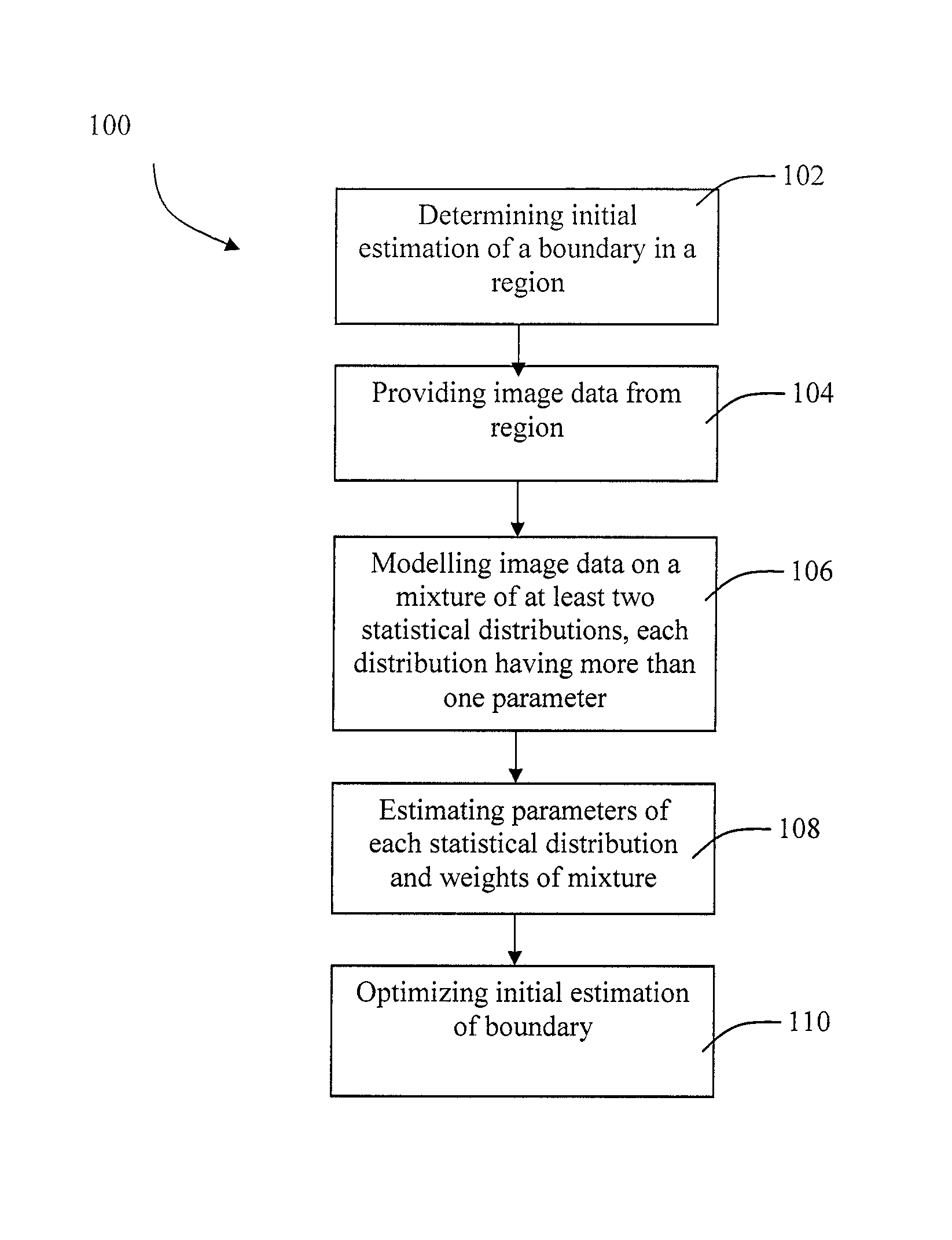
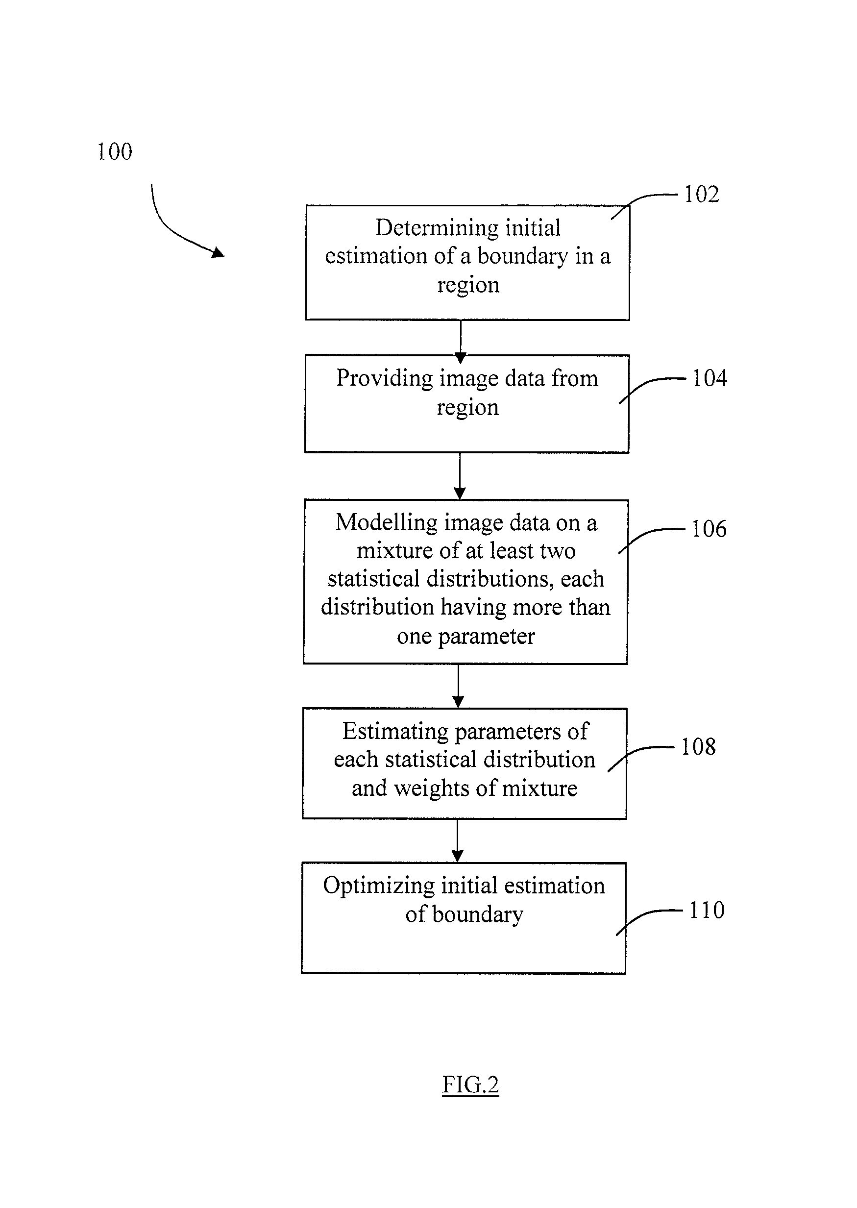
Image segmentation
a technology of image segmentation and image, applied in image enhancement, instruments, ultrasonic/sonic/infrasonic diagnostics, etc., can solve the problems of time-consuming task, variability between observers and subjective interpretation, and hampered ultrasound image diagnosis
- Summary
- Abstract
- Description
- Claims
- Application Information
AI Technical Summary
Benefits of technology
Problems solved by technology
Method used
Image

Examples
example 1
[0132]The semi-automated method of the first embodiment of the present invention (Steps 102-114) was evaluated by comparing images segmented by the semi-automated method to manually segmented images as a comparison.
[0133]Method:
[0134]N=30 video sequences of B-mode images from 15 healthy subjects were considered. It was presumed each subject had disease-free carotid arteries. For each subject, longitudinal views of the right distal common carotid artery and right proximal internal carotid were acquired by one expert radiologist, for a total of two video sequences per patient. The number of expert technicians for the manual segmentations was two.
[0135]Two versions of the method of the first embodiment were tested: 1) the MGD model (which is equivalent to a mixture of Nakagami distributions after the change of variable I=r2) estimated by the EM algorithm; 2) the Mixture of Exponential Distributions (MED) model (which is equivalent to the mixture of Rayleigh distributions after the same...
PUM
 Abstract
Description
Claims
Application Information
 - R&D Engineer
- R&D Manager
- IP Professional
- Industry Leading Data Capabilities
- Powerful AI technology
- Patent DNA Extraction
Browse by: Latest US Patents, China's latest patents, Technical Efficacy Thesaurus, Application Domain, Technology Topic, Popular Technical Reports.
© 2024 PatSnap. All rights reserved.Legal|Privacy policy|Modern Slavery Act Transparency Statement|Sitemap|About US| Contact US: help@patsnap.com