
Robust and accurate freehand 3D ultrasound

a freehand, accurate technology, applied in the field of ultrasound, can solve the problems of complicated use of ultrasound probes, additional equipment, and high implementation costs, and achieve the effects of improving ultrasound probe calibration, improving ultrasound image-guided surgical interventions, and improving accuracy

Active Publication Date: 2013-10-15
THE JOHN HOPKINS UNIV SCHOOL OF MEDICINE
View PDF7 Cites 5 Cited by
  • Summary
  • Abstract
  • Description
  • Claims
  • Application Information

AI Technical Summary

Benefits of technology

The present invention provides a method for generating 3D ultrasound imaging that improves ultrasound probe calibration for 3D ultrasound imaging, provides more robust ultrasound image registration, better provides for ultrasound image-guided surgical interventions, improves out of plane motion tracking of ultrasound speckle features, and achieves these technical effects by acquiring a first ultrasound image and a second ultrasound image, dividing them into patches, computing signal to noise ratio and skewness values for each patch, and determining if there is a plurality of good patches, computing a correlation coefficient for a corresponding pair of good patches, and constructing a 3D image using the first and second ultrasound images and the elevation distance.

Problems solved by technology

Precisely measuring the position and orientation of the ultrasound probe generally requires additional equipment, which is expensive, and complicates the use of the ultrasound probe.
The latter approach requires equipping the room with optical scanning devices, which is expensive to implement and restricts the use of the ultrasound probe to the room in which the optical scanning devices are installed.
Both of these related art solutions add considerable cost and complexity to an ultrasound system.
However, this is not so simple in the case of out of plane motion.

Method used


Image

  • Robust and accurate freehand 3D ultrasound
  • Robust and accurate freehand 3D ultrasound
  • Robust and accurate freehand 3D ultrasound

Examples


Embodiment Construction

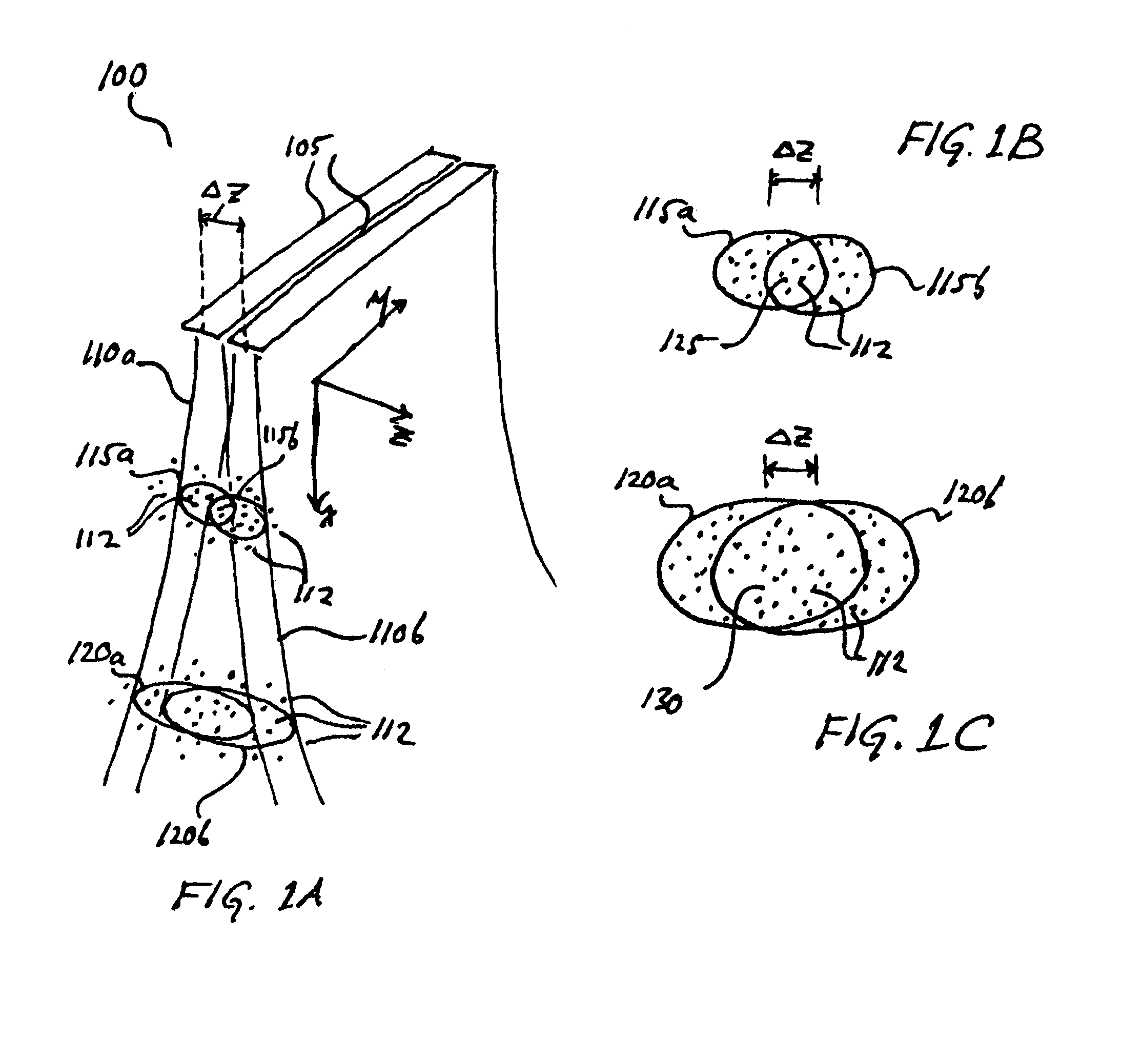
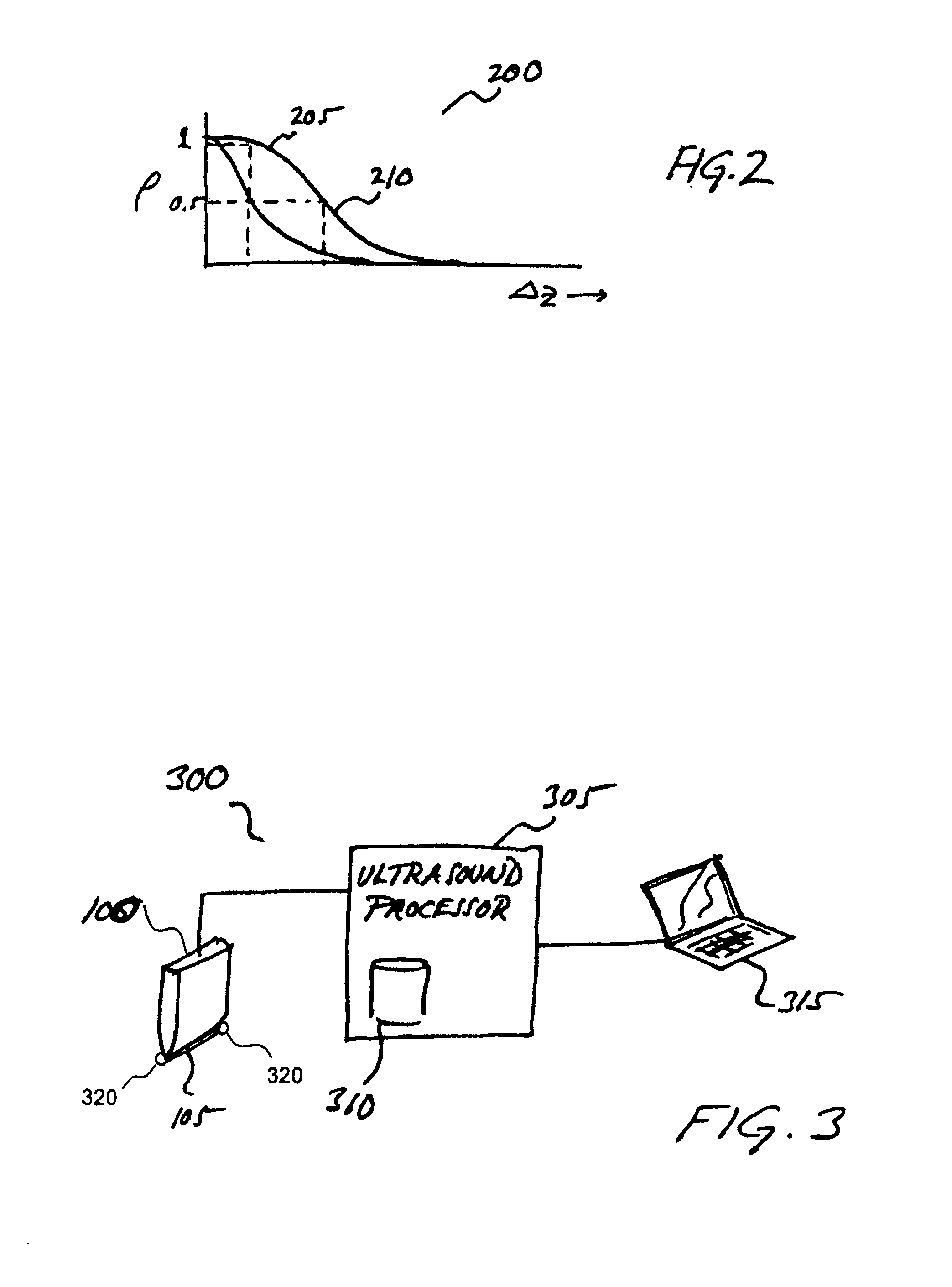
[0041]The system and method described herein exploits the traits of Fully Developed Speckle (FDS) in ultrasound imagers, whereby the extent of correlation between two images of a single FDS feature falls off at a measurable and characterizable rate as a function of out of plane distance, and as a function of depth within the ultrasound image.

[0042]FIG. 1A illustrates an ultrasound probe 100 acquiring two successive images while being scanned in an out of plane direction. Probe 100 has a transducer array 105, which is depicted at two sequential scan positions. Transducer array 105 has a first field of view 110a, corresponding to a first scan position, and a second field of view 110b, corresponding to a second scan position. First field of view 110a has a shallow resolution cell 115a and a deep resolution cell 120a. Shallow resolution cell 115a is a volume within field of view 110a that corresponds to the field of view of a single transducer for a given sample integration time. The ti...


PUM


Abstract

Disclosed is a system and method for computing out of plane motion between two ultrasound images. The method identifies regions of fully developed speckle that are common to the two images, computes a correlation coefficient corresponding to the two fully developed speckle image regions, and then computing an elevation distance corresponding to the correlation coefficient. The method exploits the measurable and characterizable relation between inter-image correlation and elevation distance, which may be determined from fully developed speckle regions. The method also identifies regions within the ultrasound images related to structure (e.g., vein or bone), and disregards these regions.

Description

[0001]This application claims the benefit of PCT Application No. PCT / US2008 / 002116, filed on Feb. 19, 2008 and U.S. Provisional Patent Application No. 60 / 901,873, filed on Feb. 16, 2007, which is hereby incorporated by reference for all purposes as if fully set forth herein.BACKGROUND[0002]1. Field of the Invention[0003]The present invention generally relates to ultrasound imaging. More particularly, the application relates to the generation of 3D images using ultrasound.[0004]2. Discussion of the Related Art[0005]Ultrasound imaging has become a widely used medical imaging modality, due in part to its effectiveness in safely imaging tissue, its ease of use, and lower cost. Ultrasound has become an essential imaging tool in applications such as identifying tissue anomalies, monitoring fetal development, and assisting in guiding surgical devices in invasive treatments.[0006]Considerable effort has recently been devoted to generating 3D images from multiple ultrasound images. By acquir...

Claims


Application Information

Patent Timeline
Patent Type & Authority Patents(United States)
IPC IPC(8): G06K9/00
CPCA61B8/483G06T7/0026G06T2207/10136G06T2207/30004A61B8/4254G06T7/32
Inventor RIVAZ, HASSANBOCTOR, EMAD MOUSSAFICHTINGER, GABORHAGER, GREGORY
Owner THE JOHN HOPKINS UNIV SCHOOL OF MEDICINE
Learn More