Radiotherapy apparatus

a technology of radiotherapy and apparatus, which is applied in the direction of diaphragms/collimeters, radiation diagnostic devices, and treatment, etc., can solve the problems of inability to switch easily, unacceptable patient skin dose, etc., and achieve the effect of reducing skin dos

Active Publication Date: 2013-01-15
ELEKTA AB
View PDF18 Cites 7 Cited by
  • Summary
  • Abstract
  • Description
  • Claims
  • Application Information

AI Technical Summary

Benefits of technology

[0013]To date, therefore, there does not appear to be a device able to switch easily between a therapeutic beam and a beam-eye-view diagnostic beam produced by the vacuum window and is therefore capable of offering good image resolution, high contrast images and low patient dose.
[0015]The present invention therefore provides a radiation source, comprising an electron gun, a pair of targets locatable in the path of a beam produced by the electron gun, the targets having different emission characteristics, and an electron absorber insertable into and withdrawable from the path of the beam. Generally, one of the targets will be thin and will also act as the vacuum window. In this way, a practical and realisable device is provided which is able to employ the principle of using the vacuum window for imaging enumerated by Galbraith in a manner that is safe and practical to use with patients. In the described embodiment, the targets are interchangeably locatable in the path, but other designs of radiation source may differ.
[0017]Thus, one target (imaging target / vacuum window) is preferably of Nickel or other suitable material (e.g. stainless steel, titanium), that can be formed into a relatively thin target, able to withstand the electron beam current, to act as a vacuum / air interface and of sufficient thickness to produce bremsstrahlung radiation but thin enough to transmit the bremsstrahlung photons so as not to be self absorbing. Such a target is capable of acting as an electron window (where desired) to allow the apparatus to produce a therapeutic electron beam. However, the atomic number properties of Nickel or other suitable material formed into a thin target will allow the production of a useful bremsstrahlung radiation. The other target (therapy target) can be a conventional x-ray target such as at least one of Copper, Tungsten, or a composite including Copper and Tungsten. The electron absorber, which is located outside the vacuum chamber and is used in conjunction with the imaging target only, preferably comprises a material of atomic number lower than the vacuum window, such as Carbon, Beryllium, Aluminium etc. In addition, the use of a diagnostic filter made of Aluminium or other suitable material can be included to reduce the skin dose in imaging mode.

Problems solved by technology

To date, therefore, there does not appear to be a device able to switch easily between a therapeutic beam and a beam-eye-view diagnostic beam produced by the vacuum window and is therefore capable of offering good image resolution, high contrast images and low patient dose.
The lower atomic number electron beam absorber serves to remove the residual electrons transmitted through the vacuum window (which otherwise would result in unacceptable levels of patient skin dose).

Method used


Image

  • Radiotherapy apparatus
  • Radiotherapy apparatus
  • Radiotherapy apparatus

Examples


Embodiment Construction

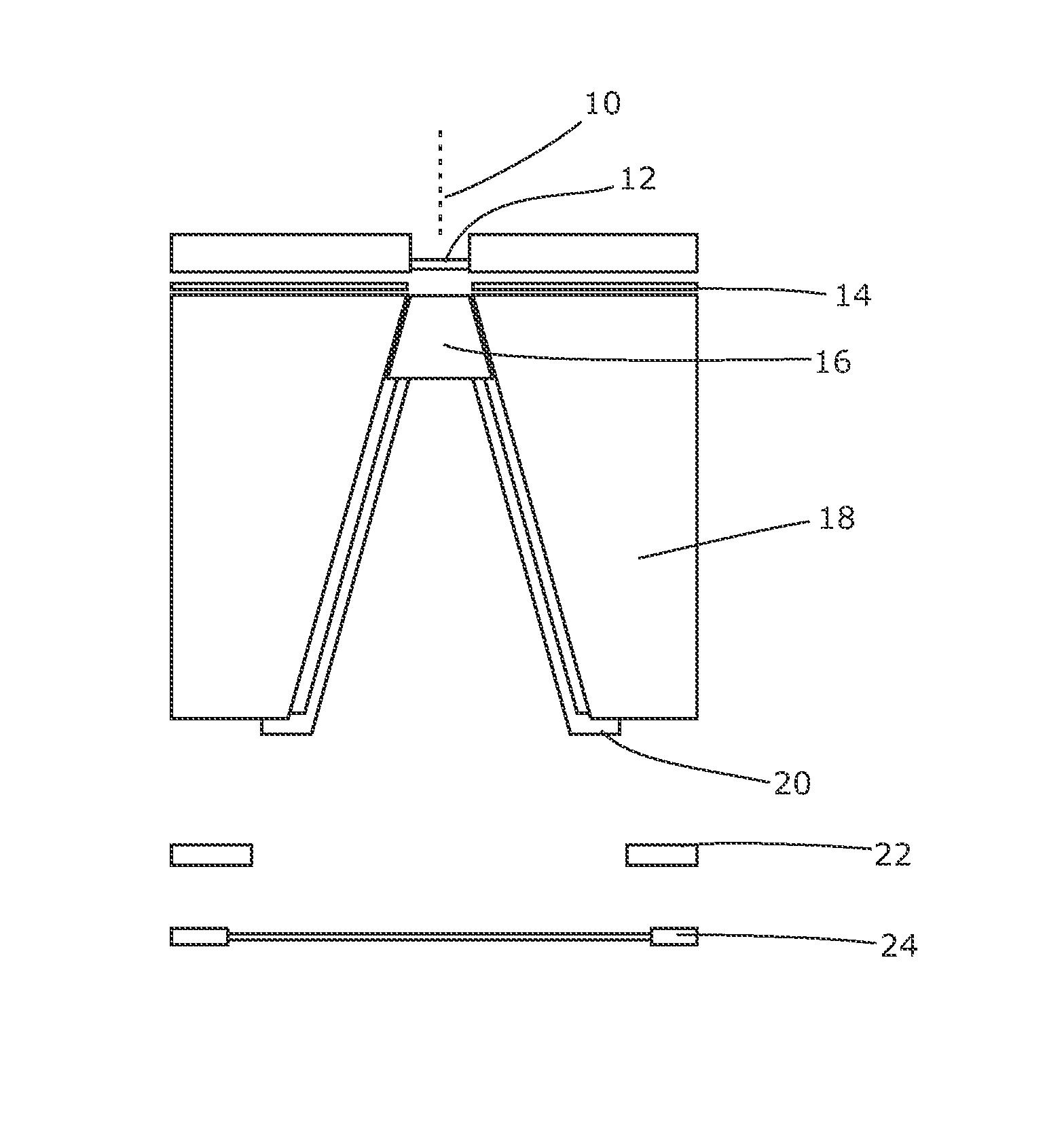
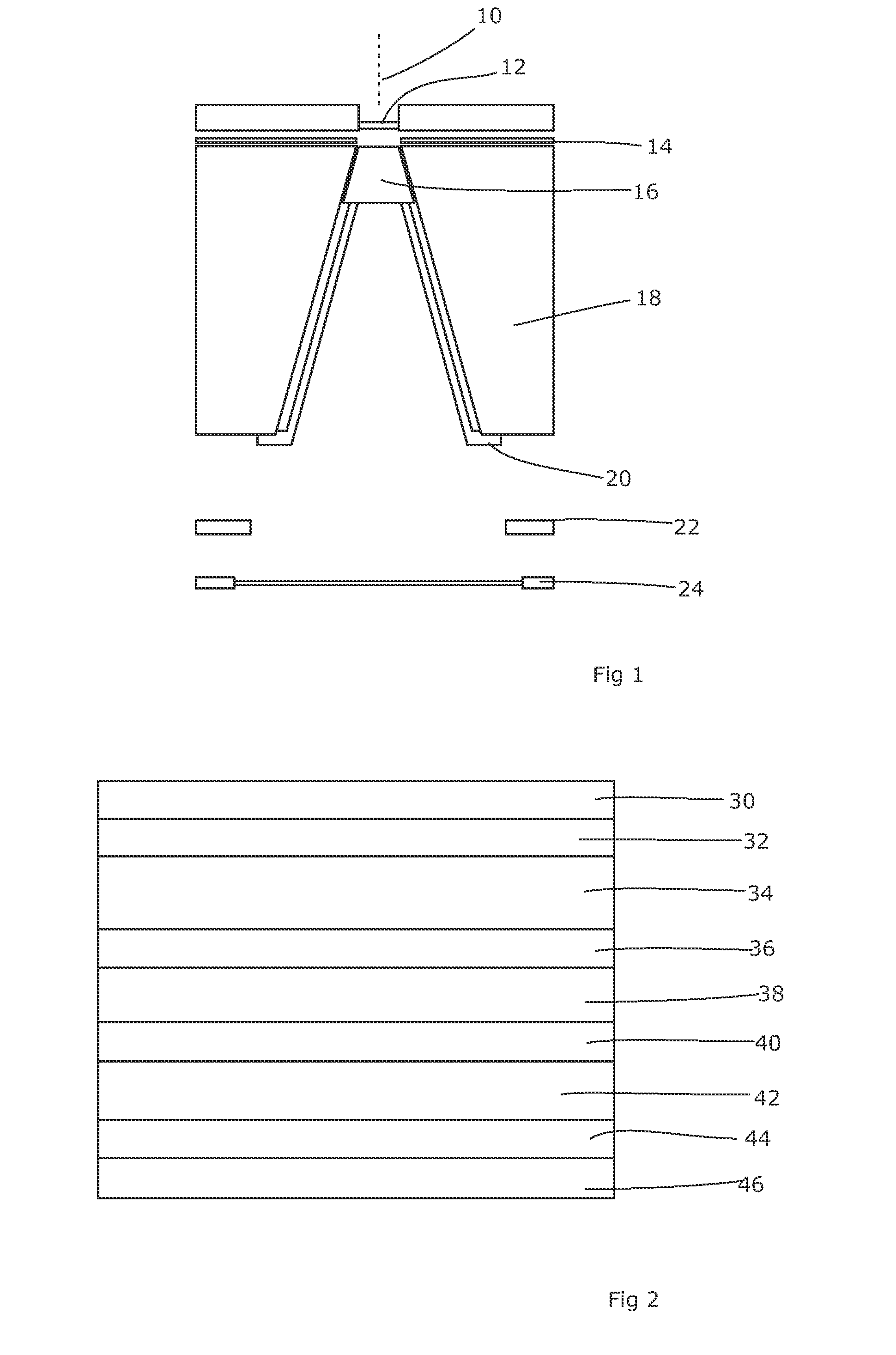
[0035]The successful treatment of cancer with radiotherapy requires a large radiation dose to be deposited accurately in both position and intensity. To verify that the patient is in the correct position, portal images have traditionally been acquired throughout the patient's treatment. These images are produced using the megavoltage treatment beam and (unfortunately) suffer from inherently low contrast. This in turn limits the ability to position the patient accurately. An increase in accuracy could potentially lead to higher tumour control and / or lower normal tissue complication with the expectation of improved therapeutic benefit.

[0036]Several methods for improving this situation have been proposed and fall into three categories. The first method involves changing the object properties, for example by inserting fiducial markers in the treatment region. Secondly, improvements to the imaging device can be made, and thirdly the imaging beam spectrum may be modified. The latter eithe...


PUM


Abstract

It is desirable to achieve a co-incident investigative kV source for a therapeutic MV source—a so-called “beams-eye-view” source. It has been suggested that bremsstrahlung radiation from an electron window be employed; we propose a practical structure for achieving this which can switch easily between a therapeutic beam and a beam-eye-view diagnostic beam capable of offering good image resolution. Such a radiation source comprises an electron gun, a pair of targets locatable in the path of a beam produced by the electron gun, one target of the pair being of a material with a lower atomic number than the other, and an electron absorber insertable into and withdrawable from the path of the beam. In a preferred form, the electron gun is within a vacuum chamber, and the pair of targets are located at a boundary of the vacuum chamber. The lower atomic number target can be Nickel and the higher atomic number target Copper and / or Tungsten. The electron absorber can be Carbon, and can be located within the primary collimator, or within one of a plurality of primary collimators interchangeably locatable in the path of the beam. Such a radiation source can be included within a radiotherapy apparatus, to which the present invention further relates. A flat panel imaging device for this source can be optimised for low energy x-rays rather than high energy; Caesium Iodide-based panels are therefore suitable.

Description

FIELD OF THE INVENTION[0001]The present invention relates to radiotherapy apparatus.BACKGROUND ART[0002]The accurate delivery of radiotherapy to a patient depends on a number of factors, including the accurate determination of the patient's current position, in terms of both their gross external position and the position of the internal structures that are to be irradiated or avoided. Some form of investigative x-ray apparatus is therefore a valuable part of a radiotherapy apparatus.[0003]Given that the apparatus itself is capable of producing a beam of x-rays, it might be thought that this could be used as an investigative source. However, the therapeutic beam is typically at a high energy (in the MV range) and therefore the image contrast is poor and the dose delivered to the patient is relatively high. The poor contrast results from the attenuation coefficients that apply at higher energies as opposed to those that apply, at lower energies. At higher energies, the coefficients of...

Claims


Application Information

Patent Timeline
Patent Type & Authority Patents(United States)
IPC IPC(8): A61N5/10H01J35/08G21K3/00
CPCA61N5/1049G21K1/10H01J35/08G21K1/02A61B6/06A61B6/4042A61N2005/1061A61B6/583H01J2235/081H01J35/116
Inventor BROWN, KEVINTHOMPSON, MARIA GIULIAHANSEN, VIBEKE NORDMARKEVANS, PHILIP MARKROBERTS, DAVID ANTHONY
Owner ELEKTA AB
Why Eureka
Try Eureka