Image analysis
a technology of image analysis and mitotic activity, applied in image analysis, instruments, computing, etc., can solve the problems of difficult to obtain the qualification to perform such examination, frequent review, and exacerbated by the complexity of some samples, and achieve cost-effective effects
- Summary
- Abstract
- Description
- Claims
- Application Information
AI Technical Summary
Benefits of technology
Problems solved by technology
Method used
Image
Examples
Embodiment Construction
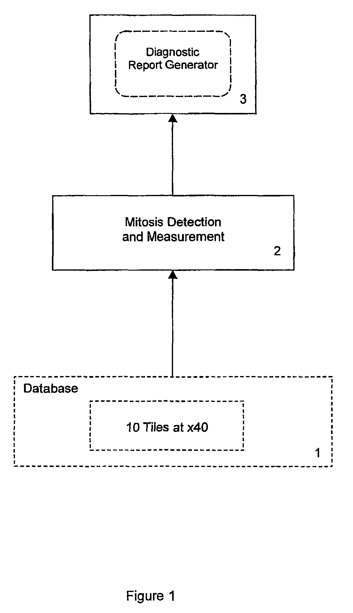
[0018]FIG. 1 shows a process for the assessment of tissue samples in the form of histopathological slides of potential carcinomas of the breast. The process measures mitotic activity of epithelial cells to produce a parameter for use by a pathologist as the basis for assessing patient diagnosis. It employs a database 1, which maintains digitised image data obtained from histological slides. Sections are cut from breast tissue samples (biopsies), placed on respective slides and stained using the staining agent Haematoxylin & Eosin (H&E), which is a common stain for delineating tissue and cellular structure.
[0019]To obtain the digitised image data for analysis, a histopathologist scans a slide under a microscope and at 40× magnification selects regions of the slide which appear to be most promising in terms of analysing mitotic activity. Each of these regions is then photographed using the microscope and a digital camera. In one example a Zeiss Axioskop microscope has been used with a...
PUM
 Abstract
Description
Claims
Application Information
 - R&D
- Intellectual Property
- Life Sciences
- Materials
- Tech Scout
- Unparalleled Data Quality
- Higher Quality Content
- 60% Fewer Hallucinations
Browse by: Latest US Patents, China's latest patents, Technical Efficacy Thesaurus, Application Domain, Technology Topic, Popular Technical Reports.
© 2025 PatSnap. All rights reserved.Legal|Privacy policy|Modern Slavery Act Transparency Statement|Sitemap|About US| Contact US: help@patsnap.com


