
Medical device and method for human tissue and foreign body extraction

a technology of human tissue and foreign body, applied in the field of medical devices and methods, can solve the problems of injury, death, and inability to fit through the small openings in the skin, and take too long to de-bulk and remove the tissue,

Active Publication Date: 2012-04-10
DAI Z
View PDF52 Cites 84 Cited by
  • Summary
  • Abstract
  • Description
  • Claims
  • Application Information

AI Technical Summary

Benefits of technology

The device enables faster and safer extraction of tissue masses with reduced risk of damage to surrounding tissue and eliminates the need for gas insufflation, improving surgical efficiency and reducing post-surgical cleanup.

Problems solved by technology

But a problem arises when that mass will not fit through the small openings in the skin.
Many skilled surgeons don't do MIS in some cases because extraction of the tissue from the patient's body would necessitate a big incision or it would take too long to de-bulk and remove the tissue.
But this design has many drawbacks.
First of all, the motorized tubular knife is exposed so that it can engage and de-bulk the tissue, but the exposed knife can also contact and cause unintentional damage to surrounding tissue resulting in injury, hospitalization, and / or death.
In addition, the tissue removal rate is dependent on the composition and stability of the tissue, the skill of the physician, the fatigue and frustration of the physician (tissue removal is typically the last step in the procedure before closing the patient), etc.
Moreover, although current morcellating devices don't necessarily require the use of an assistant during the surgery, many surgeons find the use of the devices to be so tiresome and cumbersome that they end up using an assistant anyway.

Method used


Image

  • Medical device and method for human tissue and foreign body extraction
  • Medical device and method for human tissue and foreign body extraction
  • Medical device and method for human tissue and foreign body extraction

Examples


Embodiment Construction

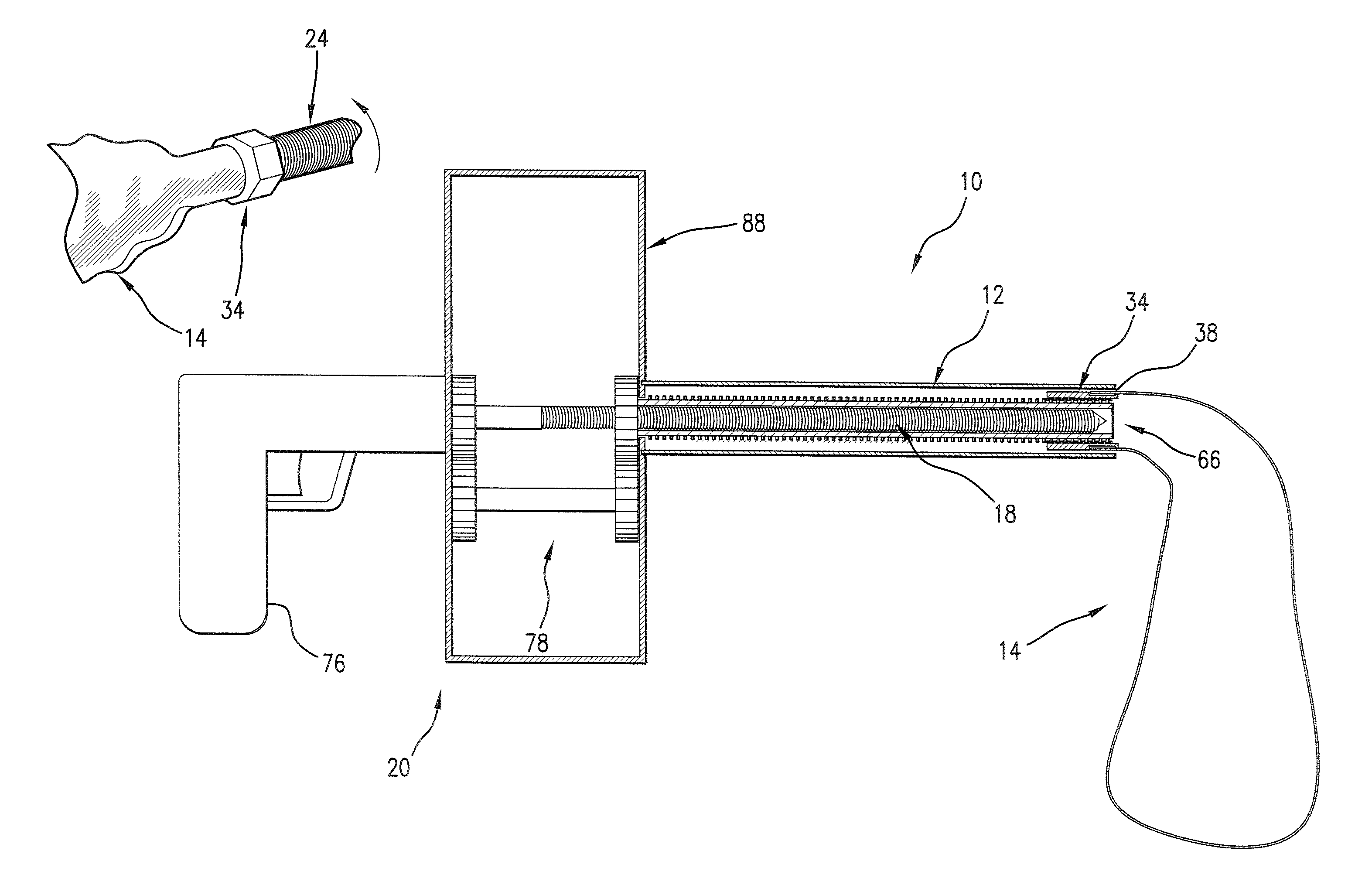
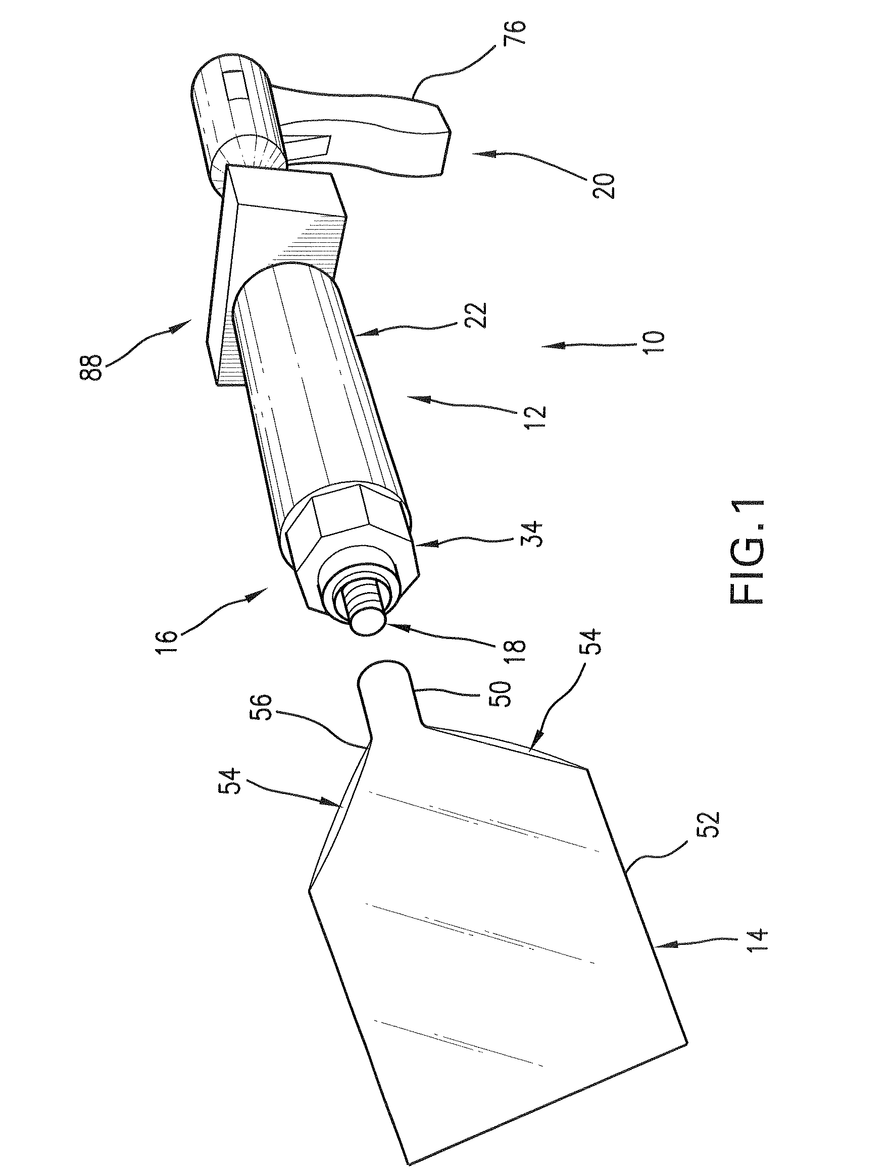
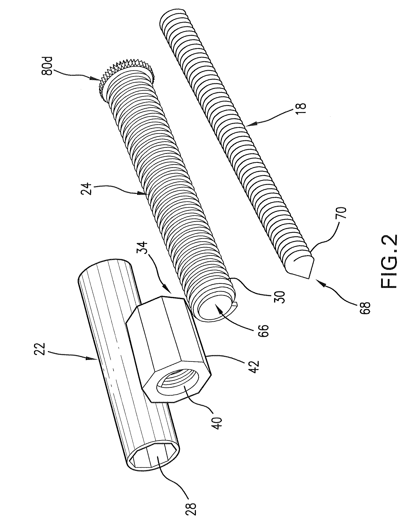
[0054]Referring now to the drawing figures, FIGS. 1-5B show a tissue capture and extraction device 10 according to an example embodiment of the present invention. The device 10 includes a coaxial tube assembly 12, a capture bag 14, a bag-translating assembly 16, a de-bulking tool 18, and a drive assembly 20. The drive assembly 20 operates the bag-translation assembly 16 to move the bag 14 through the coaxial tube assembly 12 to deliver the bag into a cavity in a patient's body. After a mass of tissue in the cavity is freed from its surroundings and placed in the bag 14, the drive assembly 20 operates the bag-translation assembly 16 to begin retracting the bag back into the coaxial tube assembly 12. As the bag 14 is being retracted, the mass in the bag is brought into engagement with the de-bulking tool 18. Then the drive assembly 20 operates the de-bulking tool 18 to morcellate the mass in the bag 14 and extract the morcellated mass bits through the coaxial tube assembly 12. The dev...


PUM


Abstract

A coaxial tube assembly, a bag, a bag-translating assembly, a de-bulking tool, and a drive assembly. The bag is delivered into a cavity in a patient's body either manually for open surgery or through the coaxial tube assembly by operation of the drive assembly for minimally invasive surgery. After a mass of tissue is placed in the bag, which is now secured to the bag-translating assembly, the drive assembly is operated to activate the bag-translation assembly to retract the bag into the annular space of the coaxial tube assembly. As the bag is being retracted, the mass in the bag is pulled into engagement with the de-bulking tool, which extends through the lumen of the coaxial tube assembly. The drive assembly activates the de-bulking tool to morcellate the mass in the bag and convey the morcellated bits of the mass through the lumen of the coaxial tube assembly.

Description

TECHNICAL FIELD[0001]The present invention relates generally to medical devices and methods and, in particular, to removal of masses such as tissue and foreign matter from the bodies of humans or other animals.BACKGROUND OF THE INVENTION[0002]Minimally Invasive Surgery (MIS) is becoming more and more prevalent and preferred by patients. This is because MIS typically results in faster patient recovery times and shorter hospital stays, and oftentimes the procedure can be done on an outpatient basis. MIS can be in the form of endoscopic, mini-incision, robotic, or natural orifice surgery, and thus is sometimes referred to as no-scar, low-scar, bandage, keyhole, or pinhole surgery. To perform MIS the surgeon uses a variety of instruments to cut, coagulate, seal, desiccate, manipulate, denature, or otherwise work on tissue. Modern technology has effectively shrunken down the size of these devices so that they can fit through very small openings in the skin. So surgeons can insert these i...

Claims


Application Information

Patent Timeline
Patent Type & Authority Patents(United States)
IPC IPC(8): A61B17/24
CPCA61B17/00234A61B2017/00287A61B2017/320024
Inventor MOHAMED, ZIADMCDONALD, TIMOTHY D.
Owner DAI Z
Learn More