
Method and apparatus for ultrasound imaging and elasticity measurement

a technology applied in the field of ultrasound imaging and elasticity measurement, can solve the problems of only providing an average elasticity value for the entire tissue being compressed, and the device has a number of limitations, and achieves the effect of enhancing the propagation trace of shear wav

Active Publication Date: 2012-04-03
THE HONG KONG POLYTECHNIC UNIV
View PDF14 Cites 11 Cited by
  • Summary
  • Abstract
  • Description
  • Claims
  • Application Information

AI Technical Summary

Problems solved by technology

However, the palpation device has a number of limitations, including the requirement of tissue boundary condition.
Also, the device can only provide an average elasticity valued for the entire tissue being compressed.

Method used


Image

  • Method and apparatus for ultrasound imaging and elasticity measurement
  • Method and apparatus for ultrasound imaging and elasticity measurement
  • Method and apparatus for ultrasound imaging and elasticity measurement

Examples


Embodiment Construction

[0020]Various embodiments of the present invention will be described in detail hereinafter with reference to the accompanying drawings.

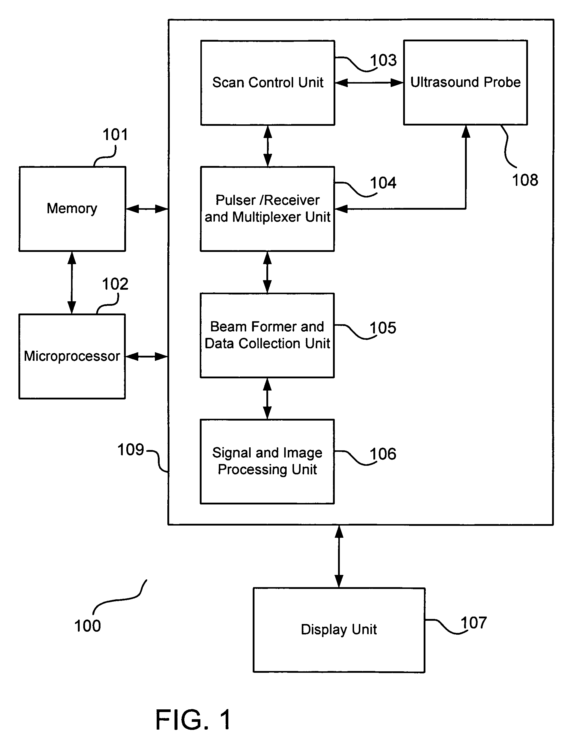
[0021]FIG. 1 is an exemplary block diagram of the hardware schematic of the present invention. Ultrasound scanning system 100 includes memory 101, microprocessor 102, ultrasound scanner 109, and display unit 107. The ultrasound scanner 109 includes a scan control unit 103, pulser / receiver and multiplexer unit 104, beam former unit 105, signal and image processing unit 106, and ultrasound probe 108. Each element in FIG. 1 is capable of communicating with another element in the ultrasound scanning system 100.

[0022]Microprocessor 102 is capable of executing instruction for controlling the ultrasound scanner 109. Microprocessor 102 may communicate with memory 101, which may include computer-executable program codes for driving the ultrasound scanner 109. The memory 101 serves as a main memory of the ultrasound system 100. The microprocessor 102 and memor...


PUM


Abstract

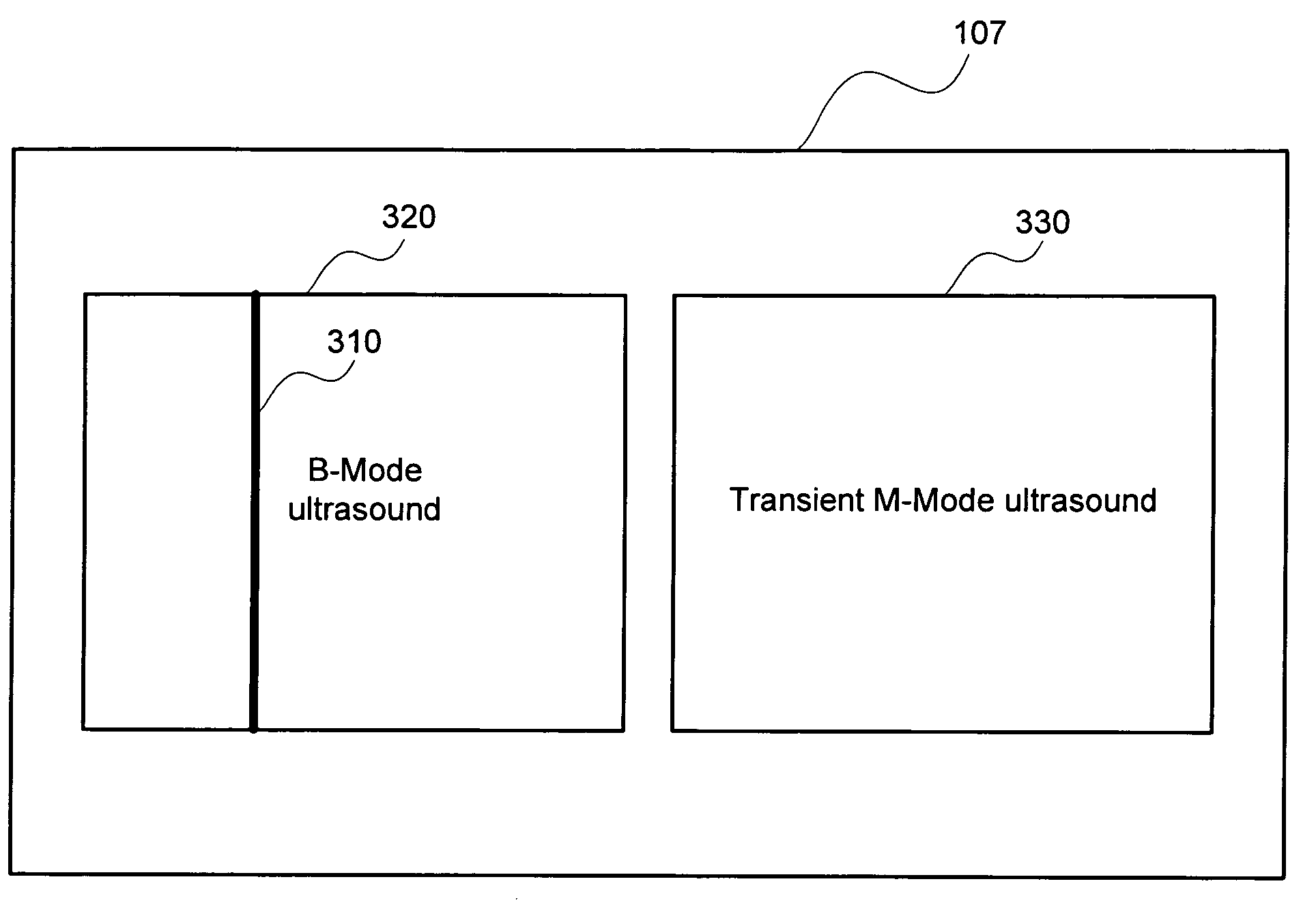
A method for performing ultrasound imaging and elasticity measurement, the method includes scanning an object to obtain a B-mode ultrasound image, selecting an A-mode signal from the B-mode ultrasound image, transmitting a high frame rate ultrasound signal, by a selected group of ultrasound transducers, at the selected A-mode signal location, forming a transient M-mode image based on the high frame rate ultrasound signal, enhancing propagation trace of shear wave based on the transient M-mode image, calculating elasticity of the object based on the propagation trace and displaying result, and displaying, simultaneously, the B-mode ultrasound image and the transient M-mode measurement result.

Description

BACKGROUND OF THE INVENTION[0001]1. Field of the Invention[0002]The present invention is related to ultrasound imaging and elasticity measurement for conventional ultrasound scanners.[0003]2. Description of the Related Art[0004]The measurement of tissue elasticity is very important in different clinical fields, such as assessment of liver fibrosis caused by various liver disease, scarring caused by burn, tissue fibrosis caused by radiotherapy, muscle stiffening caused by contraction and fatigue, etc. Consequently, various techniques for measuring tissue elasticity have been developed, for example, U.S. Pat. No. 6,494,840 teaches a tissue palpation device based on ultrasound indentation technique for quantitative measurement of tissue elasticity. By using an ultrasound imaging probe together with ultrasound indentation measurement, tissue elasticity can be measured with the guide of ultrasound imaging.[0005]However, the palpation device has a number of limitations, including the requ...

Claims


Application Information

Patent Timeline
Patent Type & Authority Patents(United States)
IPC IPC(8): A61B8/00
CPCA61B8/00A61B8/485A61B8/463
Inventor ZHENG, YONGPING
Owner THE HONG KONG POLYTECHNIC UNIV
Learn More