Substance that specifically recognizes PD-1
a substance and pd technology, applied in the field of substances, can solve the problems of many unknown matters
- Summary
- Abstract
- Description
- Claims
- Application Information
AI Technical Summary
Benefits of technology
Problems solved by technology
Method used
Image
Examples
example 1
(1) Preparation of Anti-PD-1 / Anti-TCR Hybrid Antibodies
(1-A) Introduction of Maleimide to Anti-Mouse CD3ε Monoclonal Antibodies
[0113]Anti-mouse CD3ε monoclonal antibodies were substituted with Sodium phosphate (0.1M, pH7.0) and Nacl (50 mM), then 200 times quantity of sulfo-EMCS (manufactured by Dojin Chemical) were added and incubated at 20° C. for 1 hour. Then the reaction mixture was size-fractionated by gel filtration using Sephacril S-300 [Sodium phosphate (0.1M, pH7.0)] and major peak fractions were collected by monitoring the absorbency at 280 nm. The protein content was calculated from the absorbency at 280 nm simultaneously.
(1-B) Reduction of Anti-PD-1 Antibodies
[0114]Antibodies against mouse PD-1 (produced by the hybridoma cells named (J43) and deposited as international deposition Accession number FERM BP-8118) were substituted with Sodium phosphate (0.1M, pH6.0), added with 2-mercaptoethylamine (final concentration of 10 mM) and EDTA (final concentration of 1 mM), and re...
example 2
Confirmation of Reactivity of Anti-PD-1 / Anti-TCR Hybrid Fab Antibodies on Cell Surface Antigen (PD-1 and CD3)
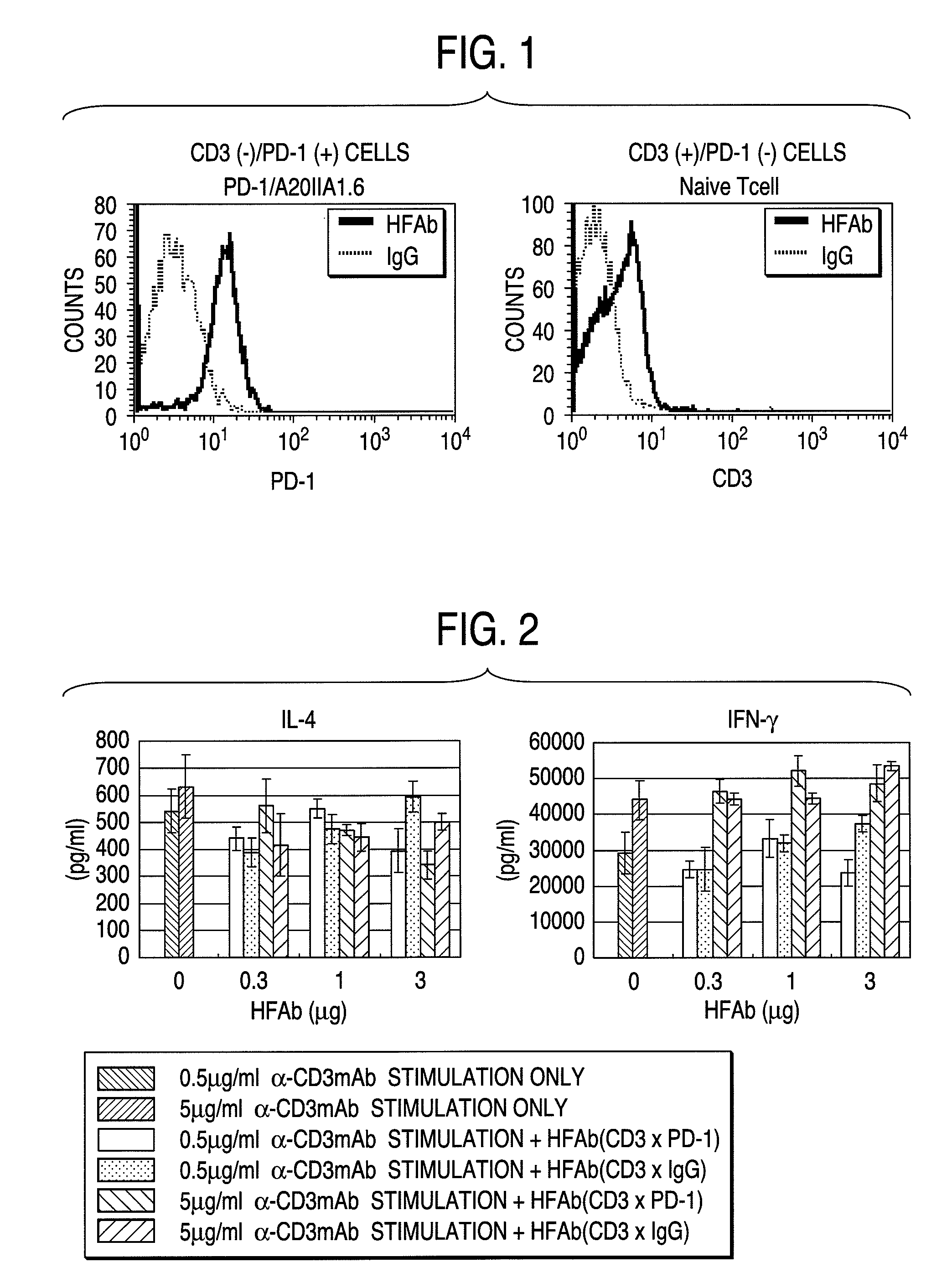
[0122]1×10⁶ cells were recovered from mPD-1 / A20IIA1.6 cells as a PD-1 positive and CD3 negative cell, and naive T cells prepared from mouse spleen cells as a PD-1 negative and CD3 positive cell, respectively. The cells were added with 91 μl of FACS buffer (PBS(−) containing 0.5% BSA, EDTA (2 mM) and 0.01% NaN3), 5 μl of mouse serum and 4 μl of hybrid antibodies (2 μg) and incubated for 30 minutes on ice. After washing with PBS(−) once, the cells were added with each 2 μl (1 μg) of second antibodies, fill upped to final 100 μl with FACS buffer, and incubated for 30 minutes on ice. Then they were analyzed by using FACScan. The results were shown in FIG. 1 (in the following figures, hybrid Fab antibody simply referred to as “HFAb”).
[0123]Analysis whether the prepared anti-PD-1 / anti-TCR hybrid Fab antibodies actually react with the cell surface antigens by using FACSsort (manufac...
example 3
Effect of Anti-PD-1 / Anti-TCR Hybrid Fab Antibodies on Activated T Cells
(A) Preparation of Spleen T Cells
[0124]Spleen was excised from BALB / c mouse. After red blood cells were hemolyzed, the spleen cells were washed once with PBS (−) and suspended in medium RPMI 1640 (10% FCS, antibiotics) (1×10⁸ cells / ml). Next, T cells were prepared by using nylon fiber column (manufactured by WAKO) for T cell separation equilibrated with the medium.
(B) Effect of Anti-PD-1 / Anti-TCR Hybrid Fab Antibodies on Activated Spleen T Cells
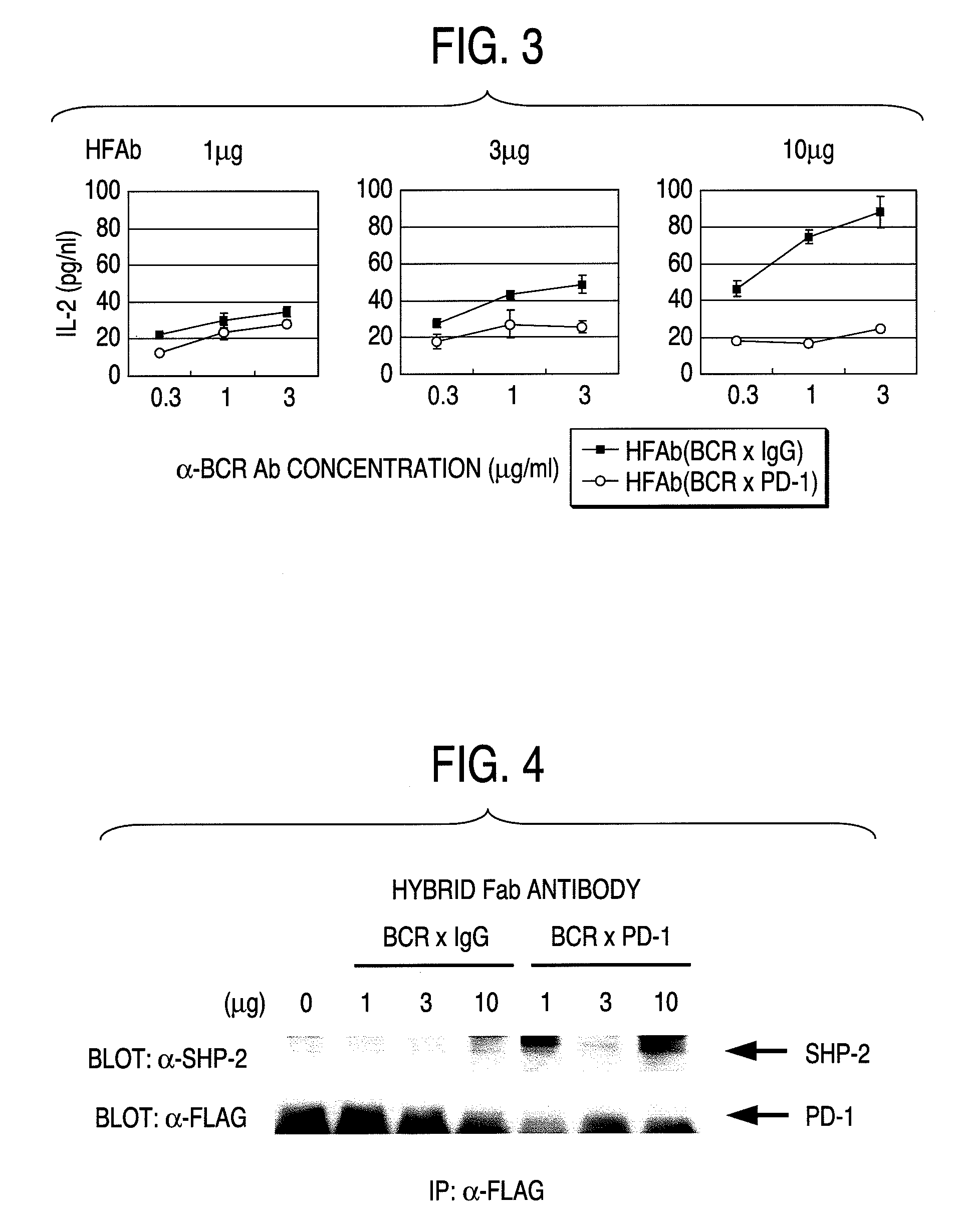
[0125]Ninety six-well plates were coated with 0.5 μg / ml and 5 μg / ml of anti-CD3 antibodies (clone KT3, manufactured by Immuntech) at 37° C. for 3 hours. T cells (2×10⁵ cells / well / 200 μl) were seeded on the plates, anti-PD-1 / anti-TCR hybrid antibodies (0.03, 0.1, 0.3, 1, 3 μg / 100 ml) were added, and incubated in a CO2 incubator (at 37° C.). After 72 hours, cytokine (IFN-r, IL-2, IL-4 and IL-10) concentrations in the recovered culture supernatants were measured by using assa...
PUM
| Property | Measurement | Unit |
|---|---|---|
| body weight | aaaaa | aaaaa |
| body weight | aaaaa | aaaaa |
| concentration | aaaaa | aaaaa |
Abstract
Description
Claims
Application Information
 - Generate Ideas
- Intellectual Property
- Life Sciences
- Materials
- Tech Scout
- Unparalleled Data Quality
- Higher Quality Content
- 60% Fewer Hallucinations
Browse by: Latest US Patents, China's latest patents, Technical Efficacy Thesaurus, Application Domain, Technology Topic, Popular Technical Reports.
© 2025 PatSnap. All rights reserved.Legal|Privacy policy|Modern Slavery Act Transparency Statement|Sitemap|About US| Contact US: help@patsnap.com


