Metastatic breast and colon cancer regulated genes
a breast and colon cancer and gene technology, applied in the direction of peptide/protein ingredients, dna/rna fragmentation, fusion polypeptide, etc., can solve the problems of difficult to predict which tumors will metastasize to other organs, difficult to distinguish between these subpopulations of patients, and difficult to chart the course of treatmen
- Summary
- Abstract
- Description
- Claims
- Application Information
AI Technical Summary
Problems solved by technology
Method used
Image
Examples
example 1
[0156]This example demonstrates identification of a differentially-expressed gene in the aggressive-invasive human breast cancer cell line MDA-MB-435.
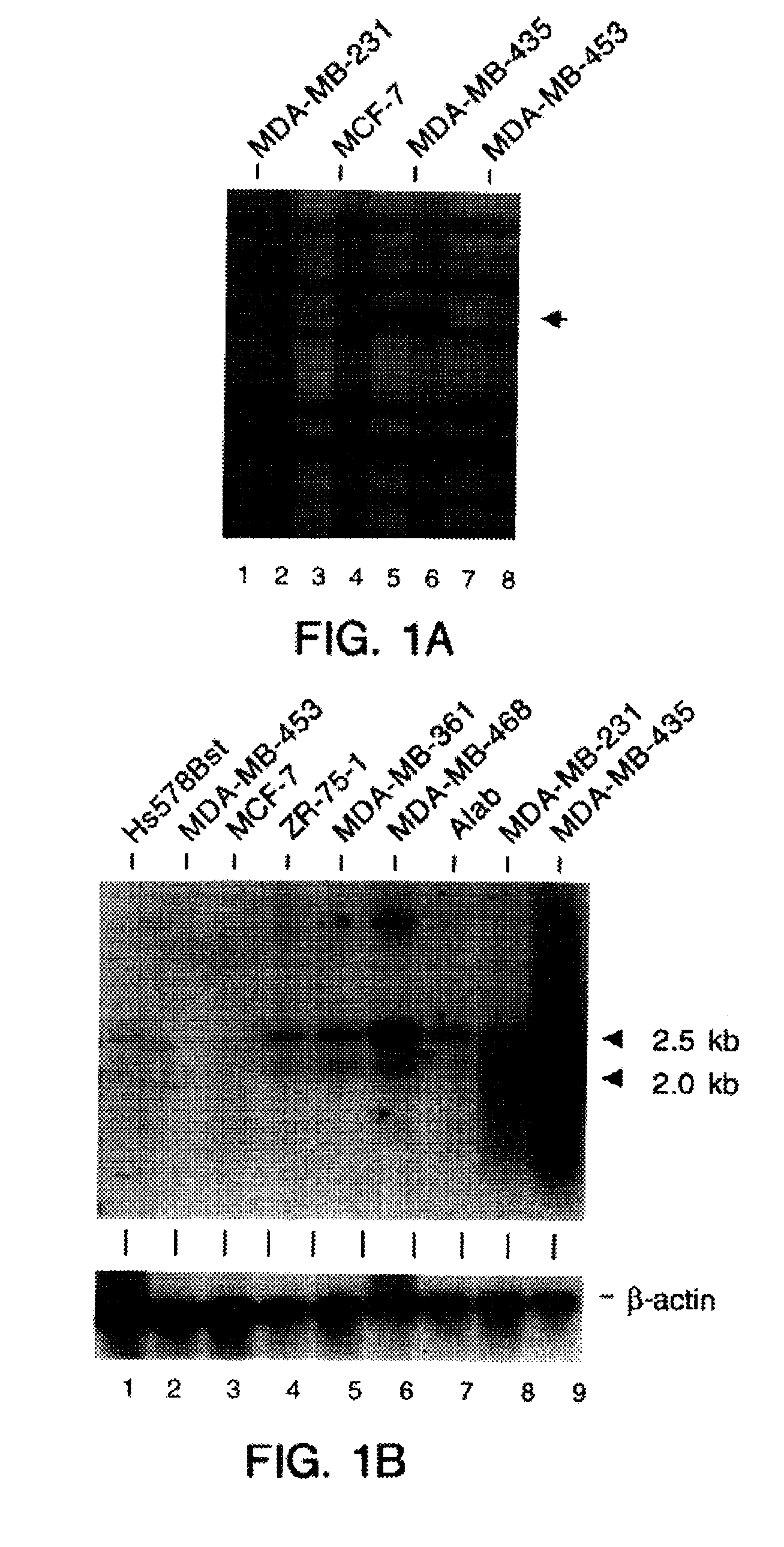
[0157]To identify genes associated with the metastatic phenotype, we compared the gene expression profiles in four human breast cancer cell lines using which display different malignant phenotypes, MDA-MB-453, MCF-7, MDA-MB-231, and MDA-MB-435, ranging from poorly-invasive to most aggressively-invasive (Engel et al., Cancer Res. 38,4327-39, 1978; Shafie and Liotta, Cancer Lett. 11, 81-87, 1990; Ozello and Sordat, Eur. J. Cancer 16, 553-59, 1980; Price et al., Cancer Res. 50, 717-21, 1990). Cell lines were chosen as starting material based on the ability to obtain high amounts of pure RNA. In contrast, human breast cancer biopsies consist of a mixture of cancer and other cell types including macrophages and lymphocytes (Kelly et al., Br. J. Cancer 57, 174-77, 1988; Whitford et al., Br. J. Cancer 62, 971-75, 1990). The described human br...
example 2
[0161]This example demonstrates the nucleotide sequence of CSP56 cDNA.
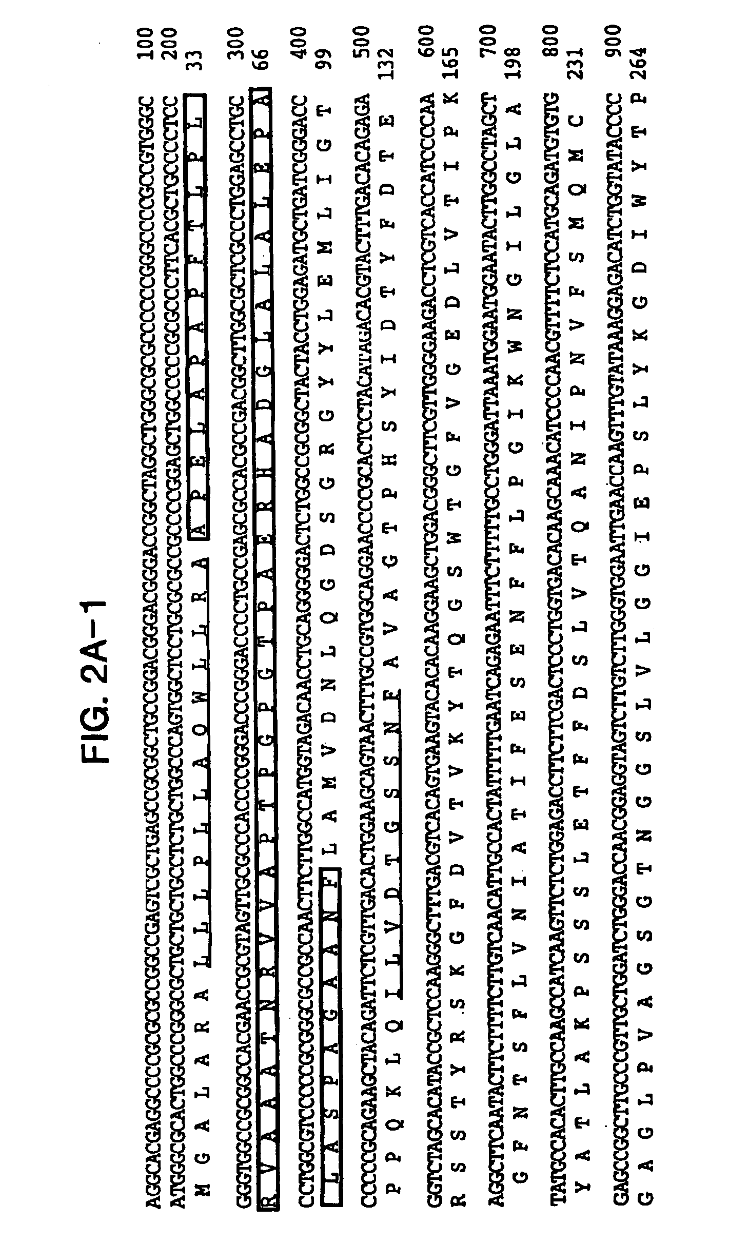
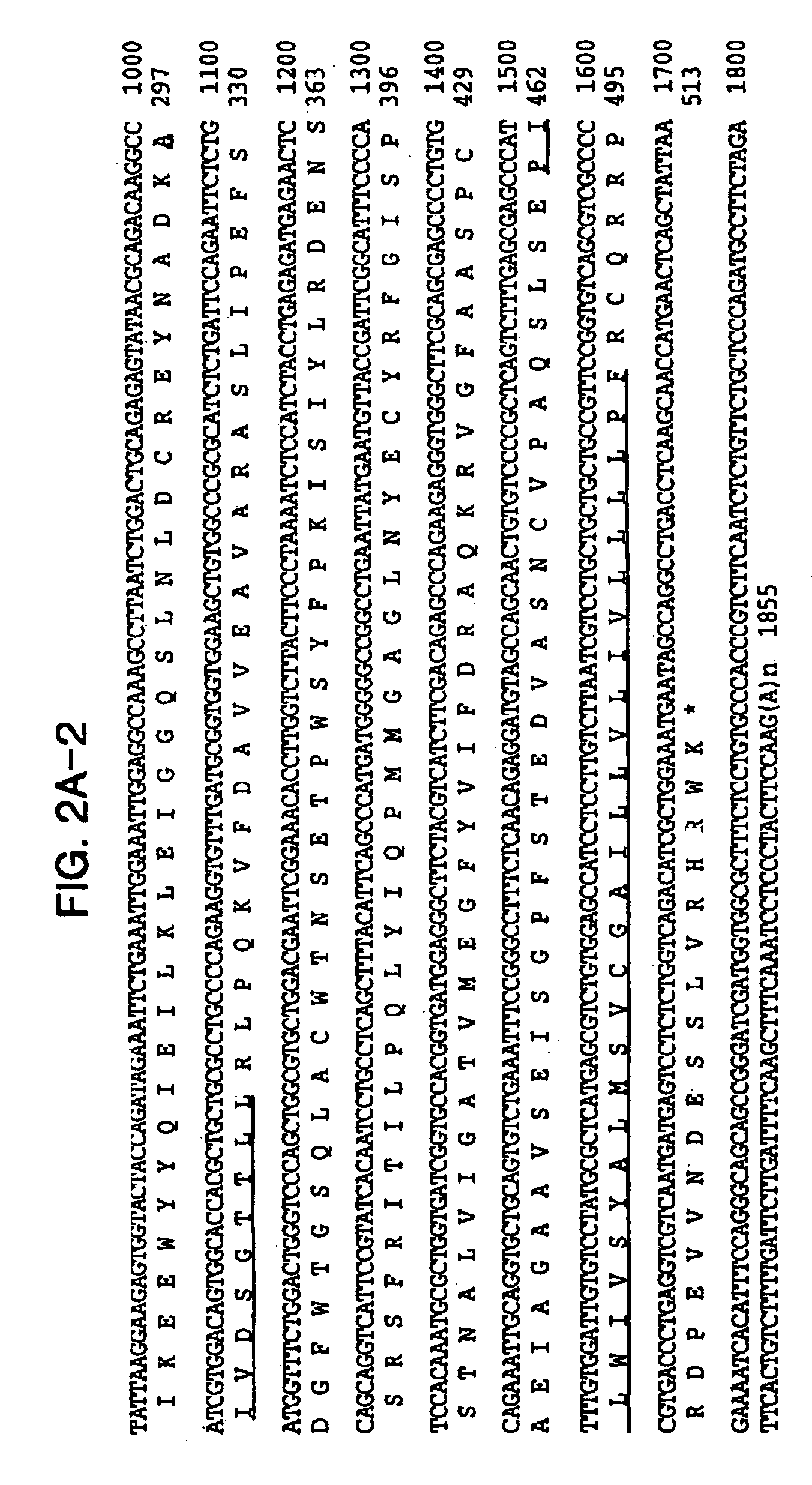
[0162]Comparison of the nucleotide sequence of CSP56 cDNA to public databases showed no significant homologies. To obtain more nucleotide sequence information, we screened a human bone marrow stromal cell cDNA library. One of the positive clones extended the original clone to 1855 nucleotides in length (FIG. 2A). This sequence was further extended at the 3′-end with several expressed sequenced tags to 2606 nucleotides in length (FIG. 2B). The additional 750 nucleotides are most probably the result of alternative poly-A site selection.
[0163]Analysis of the nucleotide sequence revealed a single open reading frame of 518 amino acids, beginning with a start codon for translation at nucleotide position 101 and terminating with a stop codon at nucleotide position 1655. A consensus Kozak sequence (Kozak, Cell 44, 283-92, 1986) around the start codon and the analysis of the codon usage (Wisconsin package, UNIX) suggests t...
example 3
[0165]This example demonstrates that CSP56 is a novel aspartyl-type protease.
[0166]Comparison of the CSP56 open reading frame with proteins in public databases shows some homology to members of the pepsin family of aspartyl proteases (FIG. 3). A characteristic feature of this protease family is the presence of two active centers which evolved by gene duplication (Davies, Ann. Rev. Biophys. Biochem. 19, 189-215, 1990; Neil and Barrett, Meth. Enz. 248, 105-80, 1995). The amino acid residues comprising the catalytic domains (Asp-Thr / Ser-Gly) and the flanking residues display the highest conservation in this family and are conserved in CSP56 (FIGS. 2 and 3).
[0167]CSP56, however, shows structural features which are distinct from other aspartyl proteases. Overall similarities of CSP56 to pepsinogen C and A, renin, and cathepsin D and E are only 55, 51, 54, 52, and 51%, respectively, neglecting the CSP56 C-terminal extension. The cysteine residues found following and preceding the catalyti...
PUM
| Property | Measurement | Unit |
|---|---|---|
| Tm | aaaaa | aaaaa |
| Tm | aaaaa | aaaaa |
| diameter | aaaaa | aaaaa |
Abstract
Description
Claims
Application Information
 - R&D
- Intellectual Property
- Life Sciences
- Materials
- Tech Scout
- Unparalleled Data Quality
- Higher Quality Content
- 60% Fewer Hallucinations
Browse by: Latest US Patents, China's latest patents, Technical Efficacy Thesaurus, Application Domain, Technology Topic, Popular Technical Reports.
© 2025 PatSnap. All rights reserved.Legal|Privacy policy|Modern Slavery Act Transparency Statement|Sitemap|About US| Contact US: help@patsnap.com


