
Method of navigating medical devices in the presence of radiopaque material

a technology of radiopaque materials and medical devices, applied in medical science, surgery, diagnostics, etc., can solve the problems of difficult to quickly and accurately navigate difficult to discern the distal end of the medical device, so as to facilitate the navigation accurately track the position of the distal end. , the effect of improving the indication of the distal end

Inactive Publication Date: 2007-07-24
STEREOTAXIS
View PDF17 Cites 242 Cited by
  • Summary
  • Abstract
  • Description
  • Claims
  • Application Information

AI Technical Summary

Benefits of technology

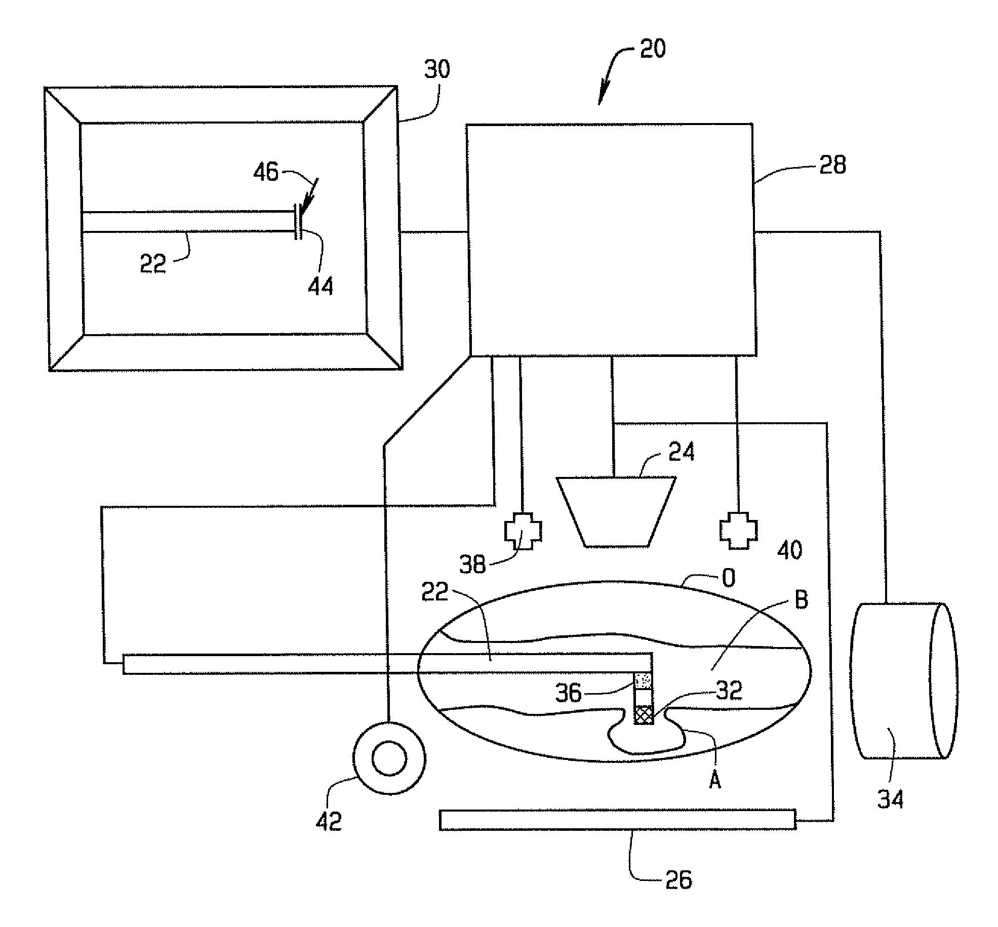
[0003]Broadly, the method of this invention relates the to navigation of medical devices in a subject's body. The method broadly comprises displaying an x-ray image of the operating region, including the distal end of the medical device; determining the location of the distal end of the medical device in a reference frame translatable to the displayed x-ray image; and displaying an enhanced indication of the distal end of the medical device on the x-ray image to facilitate the navigation of the distal end of the device in the operating region. The method of this invention allows a physician to more accurately track the position of the distal end of the medical device during navigation, this facilitates navigation, and particularly the accurate delivery of radiopaque materials in a subject's body.

Problems solved by technology

However, when there is radiopaque material in the operating region, it is often difficult to discern the distal end of the medical device.
The presence of these materials in the medical device or in the operating region can make it difficult for the physician to “see” the distal end of the medical device on x-ray images, and thus make it difficult to quickly and accurately navigate the distal end of the medical device.

Method used


Image

  • Method of navigating medical devices in the presence of radiopaque material
  • Method of navigating medical devices in the presence of radiopaque material
  • Method of navigating medical devices in the presence of radiopaque material

Examples


Embodiment Construction

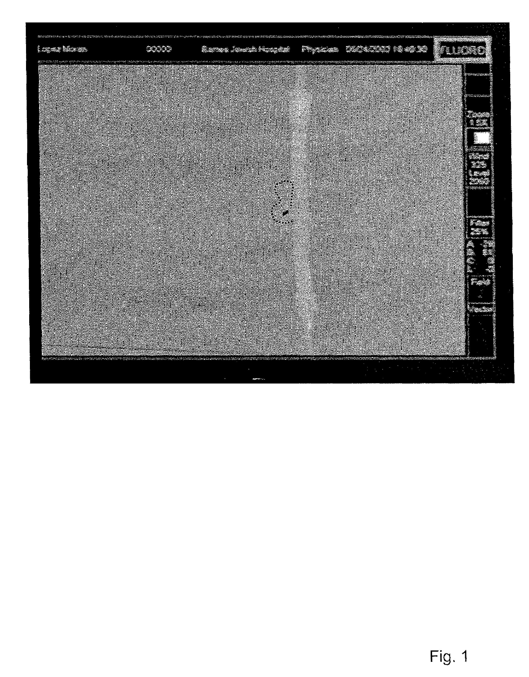
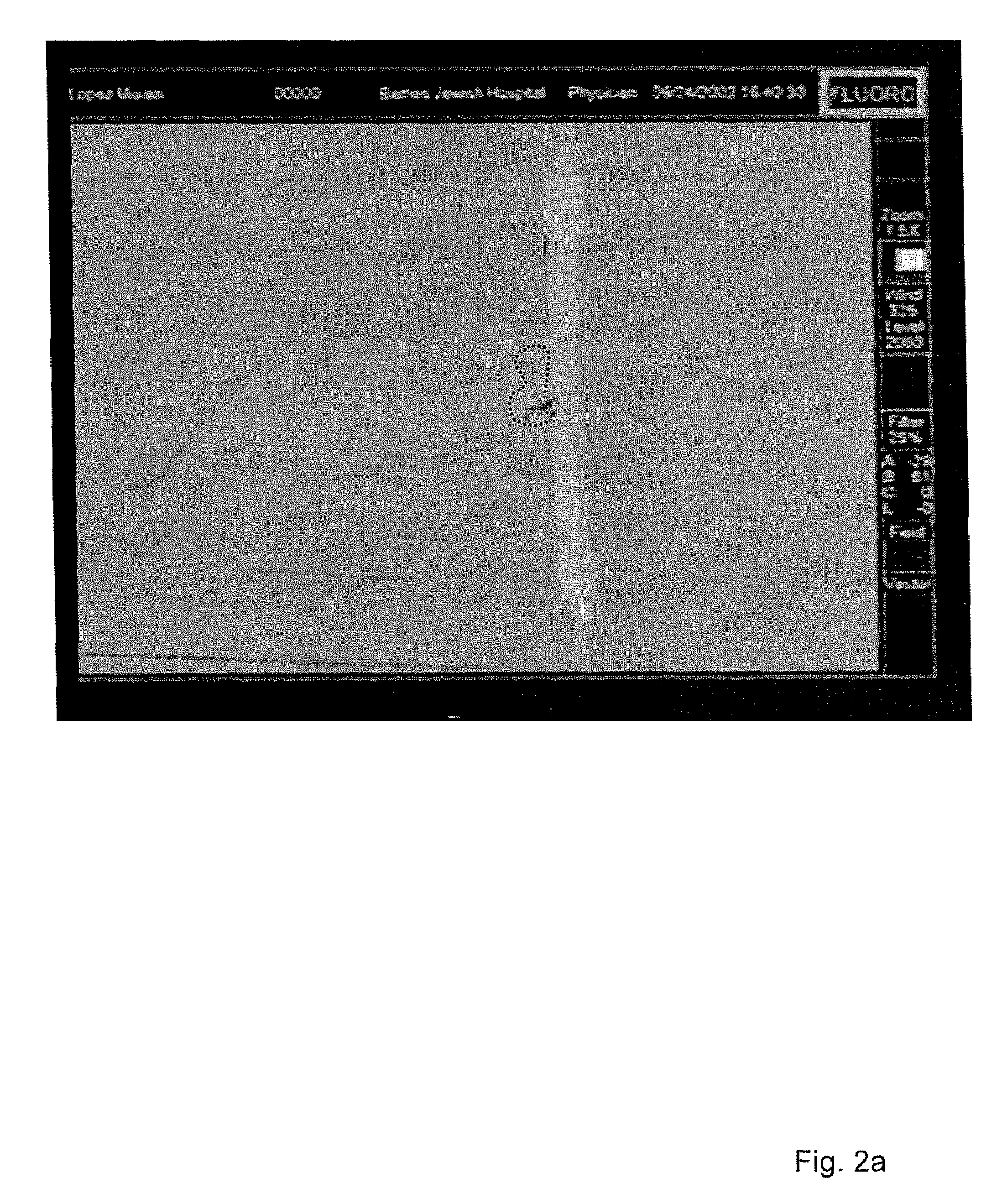
[0013]The present invention relates to a method of navigating the distal end of a medical device through an operating region in a subject's body. Broadly, this method comprises displaying an x-ray or other image of the operating region, including the distal end of the medical device, for example as shown in FIG. 1. The location of the distal end of the medical device is then determined in a reference frame translatable to the displayed x-ray image. Based upon the determined location, an enhanced indication of the distal end of the medical device is then displayed on the x-ray image to facilitate the navigation of the distal end of the device in the operating region. This is shown in FIG. 2. This helps the user identify the location of the distal end of the medical device, to facilitate navigating the device.

[0014]This method can be used with any type of medical device, such as a catheter, endoscope, guide wire, sampling (e.g. biopsy) device, drug or device delivery catheter, sensing...


PUM


Abstract

A method of navigating the distal end of a medical device through an operating region in a subject's body includes displaying an x-ray image of the operating region, including the distal end of the medical device; determining the location of the distal end of the medical device in a reference frame translatable to the displayed x-ray image; and displaying an enhanced indication of the distal end of the medical device on the x-ray image to facilitate the navigation of the distal end of the device in the operating region.

Description

FIELD OF THE INVENTION[0001]This invention relates to the navigation of medical devices in the presence of radiopaque materials, and in particular to a method of navigating a medical device for the delivery of radiopaque materials.BACKGROUND OF THE INVENTION[0002]The navigation of the distal end of a medical device through the body, whether by conventional mechanical means or magnetic means, is usually facilitated by x-ray imaging of the operating region. However, when there is radiopaque material in the operating region, it is often difficult to discern the distal end of the medical device. This occurs, for example when the medical device is delivering a radiopaque material, such as a radiopaque embolic material, or a radiopaque embolization coil. The presence of these materials in the medical device or in the operating region can make it difficult for the physician to “see” the distal end of the medical device on x-ray images, and thus make it difficult to quickly and accurately n...

Claims


Application Information

Patent Timeline
IPC IPC(8): A61B5/05A61B5/06A61B6/12
CPCA61B5/06A61B6/12A61B5/062
Inventor HASTINGS, ROGER N.FERRY, STEVEN J.LOPES, DEMETRIUS K.
Owner STEREOTAXIS
Learn More