
Device and method for modifying the shape of a body organ

a body organ and device technology, applied in the field of medical devices, can solve the problems of reducing circulatory efficiency, requiring correction, rapid deterioration, and high invasiveness of each of these procedures, and achieve the effect of reducing the chance of thrombosis formation and low metal coverage area

Inactive Publication Date: 2005-11-01
CARDIAC DIMENSIONS
View PDF128 Cites 199 Cited by
  • Summary
  • Abstract
  • Description
  • Claims
  • Application Information

AI Technical Summary

Benefits of technology

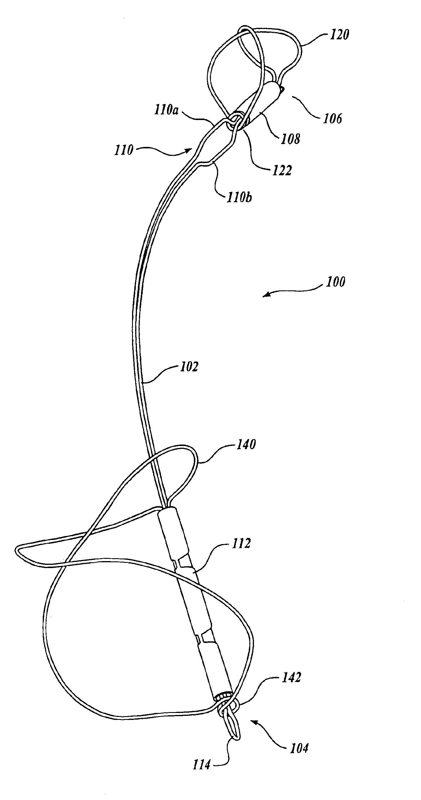
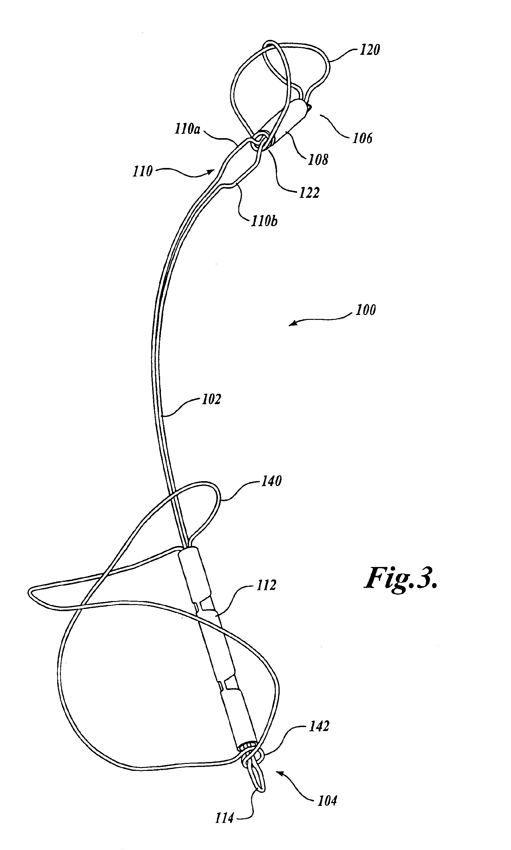
[0008]In one embodiment of the invention, the proximal and distal supports are made from a wire hoop that presents a low metal coverage area to blood flowing within the vessel. The wire hoops may allow tissue to grow over the anchors to reduce the chance of thrombosis formation. The wire hoops have a figure eight configuration and can expand to maintain contact with the vessel walls if no vessel expands or changes shape.
[0009]In another embodiment of the invention, the proximal and distal anchors of the intravascular support are rotationally offset from each other. Locks on the support wire allow a physician to ensure that the anchors have been successfully deployed and prevent the support wire from collapsing within a vessel.

Problems solved by technology

Regurgitation is a serious, often rapidly deteriorating, condition that reduces circulatory efficiency and must be corrected.
Each of these procedures is highly invasive because access to the heart is obtained through an opening in the patient's chest.
Patients with mitral valve regurgitation are often relatively frail thereby increasing the risks associated with such an operation.
While this technique appears promising, some proposed supports appear to limit the amount of blood that can flow through the coronary sinus and may contribute to the formation of thrombosis in the vessel.
Therefore, there is a need for a tissue support structure that does not inhibit the flow of blood in the vessel in which it is placed and reduces the likelihood of thrombosis formation.

Method used


Image

  • Device and method for modifying the shape of a body organ
  • Device and method for modifying the shape of a body organ
  • Device and method for modifying the shape of a body organ

Examples


Embodiment Construction

[0029]As indicated above, the present invention is a medical device that supports or changes the shape of tissue that is adjacent a vessel in which the device is placed. The present invention can be used in any location in the body where the tissue needing support is located near a vessel in which the device can be deployed. The present invention is particularly useful in supporting a mitral valve in an area adjacent a coronary sinus and vessel. Therefore, although the embodiments of the invention described are designed to support a mitral valve, those skilled in the art will appreciate that the invention is not limited to use in supporting a mitral valve.

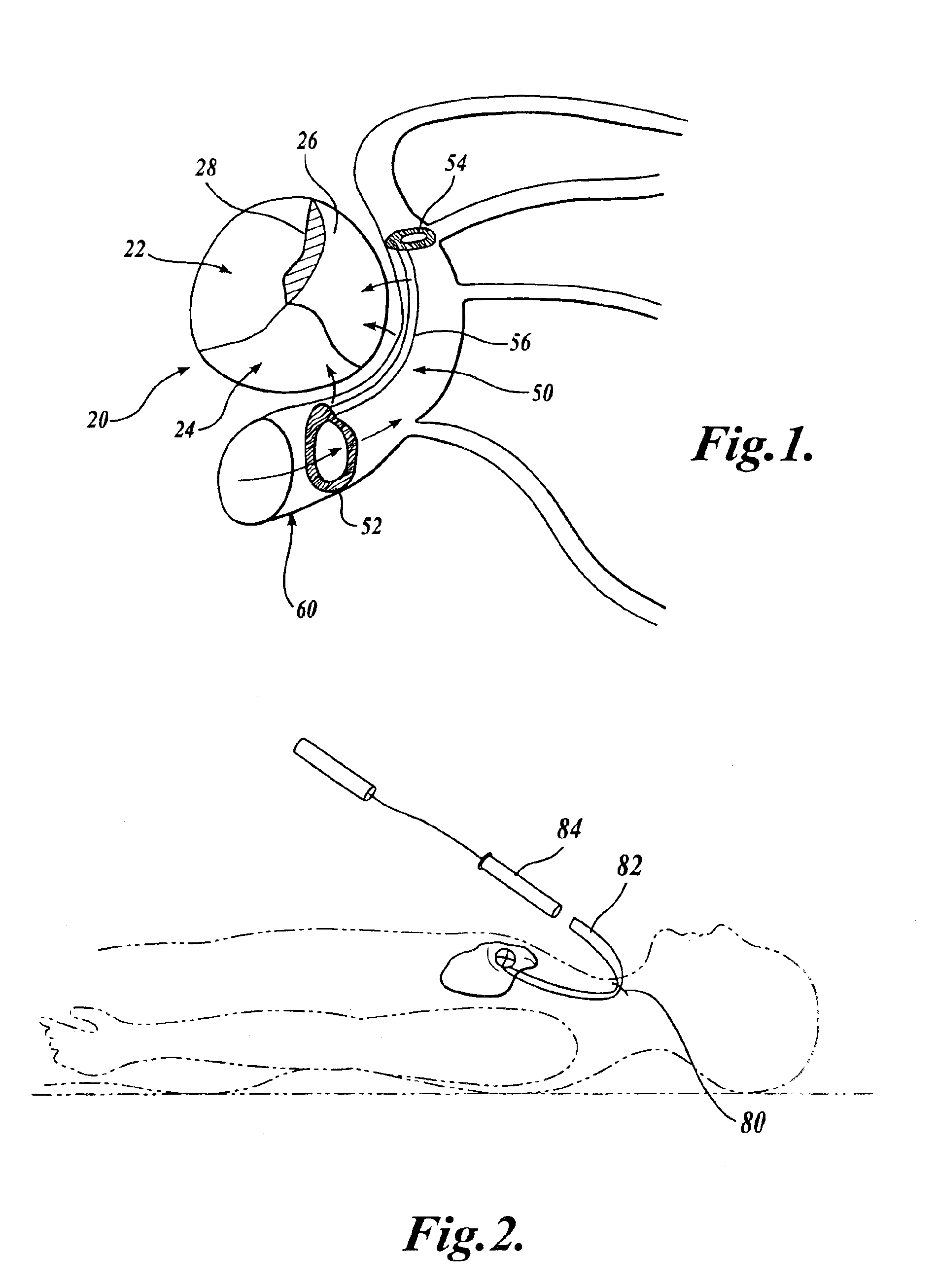
[0030]FIG. 1 illustrates a mitral valve 20 having a number of flaps 22, 24 and 26 that should overlap and close when the ventricle of the heart contracts. As indicated above, some hearts may have a mitral valve that fails to close properly thereby creating one or more gaps 28 that allow blood to be pumped back into the left atrium ...


PUM


Abstract

An intravascular support device includes a support or reshaper wire, a proximal anchor and a distal anchor. The support wire engages a vessel wall to change the shape of tissue adjacent the vessel in which the intravascular support is placed. The anchors and support wire are designed such that the vessel in which the support is placed remains open and can be accessed by other devices if necessary. The device provides a minimal metal surface area to blood flowing within the vessel to limit the creation of thrombosis. The anchors can be locked in place to secure the support within the vessel.

Description

CROSS-REFERENCES TO RELATED APPLICATIONS[0001]The present application is a continuation-in-part of U.S. application Ser. No. 10 / 331,343 filed Dec. 26, 2002, now U.S. Pat. No. 6,793,673; U.S. application Ser. No. 10 / 142,637 filed May 8, 2002, now U.S. Pat. No. 6,824,562 and U.S. application Ser. No. 10 / 066,426 filed Jan. 30, 2002, the benefit of the filing dates being claimed under 35 U.S.C. § 120 and the disclosures of which are incorporated by reference.FIELD OF THE INVENTION[0002]The present invention relates to medical devices in general, and in particular to devices for supporting internal body organs.BACKGROUND OF THE INVENTION[0003]The mitral valve is a portion of the heart that is located between the chambers of the left atrium and the left ventricle. When the left ventricle contracts to pump blood throughout the body, the mitral valve closes to prevent the blood being pumped back into the left atrium. In some patients, whether due to genetic malformation, disease or injury, ...

Claims


Application Information

Patent Timeline
IPC IPC(8): A61F2/24A61F2/06
CPCA61F2/2451
Inventor MATHIS, MARK L.NIEMINEN, GREGORY D.REUTER, DAVID G.
Owner CARDIAC DIMENSIONS
Learn More