
Conditioning protocols and use of same for tissue regeneration

a tissue regeneration and protocol technology, applied in the direction of artificial cell constructs, drug compositions, unknown materials, etc., can solve the problems of inability to meet the needs of patients, inability to achieve satisfactory/optimal treatments, and only slightly improve the quality of life of patients in need. , the treatment modality suffers from considerable disadvantages

Pending Publication Date: 2020-09-17
YEDA RES & DEV CO LTD
View PDF3 Cites 0 Cited by
  • Summary
  • Abstract
  • Description
  • Claims
  • Application Information

AI Technical Summary

Benefits of technology

The present invention provides a method for conditioning a subject in need of transplantation of progenitor cells in suspension of a tissue of interest. The method involves inducing damage to the tissue of interest by administering an agent capable of inducing damage, followed by ablating the resident stem cells in the tissue. This results in the conditioning of the subject, which can then be followed by transplanting the progenitor cells to the subject. The method can be used for the treatment of various diseases, such as cystic fibrosis, emphysema, asbestosis, chronic obstructive pulmonary disease, pulmonary hypertension, lung cancer, sarcoidosis, and others. The progenitor cells can be obtained from various sources, such as cardiac, hepatic, pancreatic, brain, nephric, and others. The method can also involve the use of herbal remedies, chemicals, antibiotics, therapeutic drugs, toxins, surgical interventions, and other methods of inducing damage to the tissue of interest.

Problems solved by technology

Pancreatic diseases such as diabetes, pulmonary diseases such as COPD, cystic fibrosis, emphysema, pulmonary fibrosis or pulmonary hypertension, liver diseases such as liver cirrhosis or viral liver diseases (e.g. Hepatitis B or C), heart diseases such as heart failure, and kidney diseases such as kidney failure are diseases of great medical and economic impact for which no satisfactory / optimal treatments are available.
Most currently available therapies only slightly improve the quality of life for the patients in need.
At present, the only definitive treatment for end-stage lung disease, liver disease, heart disease and kidney disease is the replacement of the damaged organ, but many patients die while on the waiting list due to a severe shortage of organs for transplantation and the limit for inscription being often set at 60 years of age.
Such a treatment modality, however, suffers from considerable disadvantages.
Allogeneic transplantation of organs / tissues is impossible to implement in a great many cases due to the lack of organ donors (e.g. usually cadavers) and the unavailability of suitable immunologically matched organ donors.
Furthermore, the use of living human donors often presents health risks and ethical dilemmas.

Method used


Image

  • Conditioning protocols and use of same for tissue regeneration
  • Conditioning protocols and use of same for tissue regeneration
  • Conditioning protocols and use of same for tissue regeneration

Examples


example 1

Canalicular ‘Window’ for Human and Mouse Lung Progenitors

Growth Potential of Human Embryonic Lung Tissues Harvested at Different Gestational Time Points

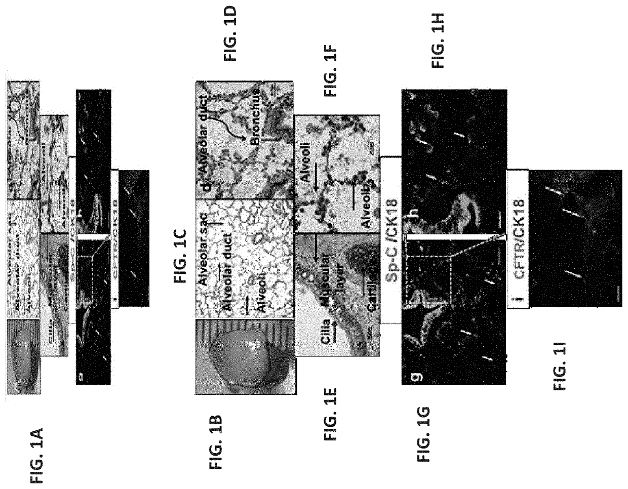
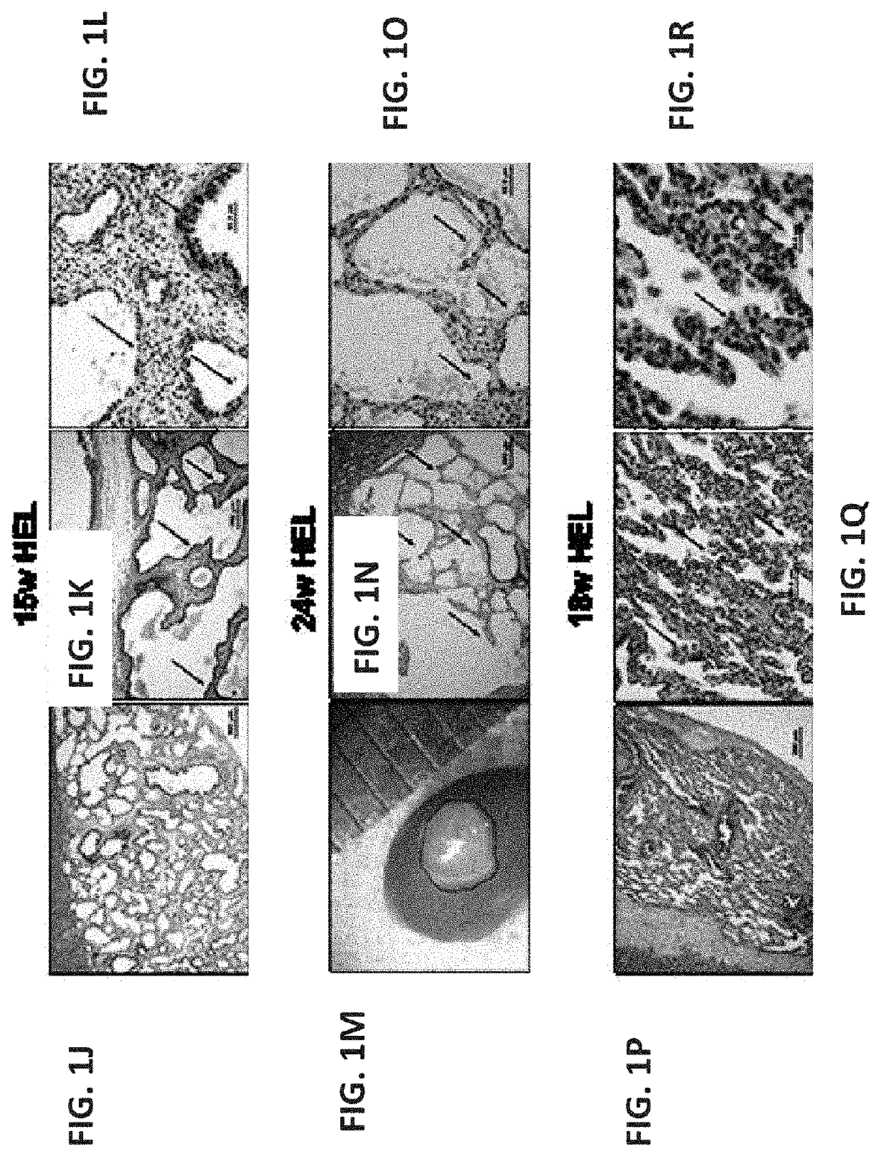
[0391]To evaluate a suitable window for human fetal lung progenitors, the present inventors initially characterized the growth and differentiation potential of lung precursor tissues harvested at different gestational time points (tissues originating from 15- to 24-week human fetuses). Using implantation of whole human (FIGS. 1A-I, FIGS. 26A-B and FIGS. 27A-L) or mouse (data not shown) fetal lung fragments under the renal capsule of immune-deficient (NOD-SCID) or syngeneic mice, respectively, it was found that use of human or mouse lung fetal tissue harvested at the canalicular stage of development enabled optimal growth and differentiation.

[0392]Overall, upon examination at 8 weeks post transplant, more than 98% of the grafts from donor tissue of all ages survived and all recovered grafts demonstrated increased size, with no evidenc...

example 2

Proof of Concept in Mouse Models for the Regenerative Potential of ‘Window’ Embryonic Lung Transplants

Optimal ‘Window’ for Harvesting Mouse Embryonic Lung Precursor Tissue for Transplantation

[0405]In order to assess the curative potential of embryonic lung derived tissue in appropriate mouse models, the optimal “window” for harvesting mouse embryonic lung for transplantation was initially defined, as for its human counterpart. Thus, mouse lung embryonic tissue was harvested at different gestational time points (E14-E17), implanted under the kidney capsule of syngeneic mice, and 8 weeks after transplantation, the implants were assessed for the presence of lung parenchyma, bronchial and alveolar structures, as well as for unwanted presence of fibrosis and cysts.

[0406]As can be seen in FIGS. 10A-E, twelve weeks after sub-capsular renal transplantation, E14 and E17 lung tissue resulted in formation of cystic and fibrotic tissue (FIGS. 10A-B), while E15-E16 mouse embryonic lung exhibited...

example 3

Ablation of Endogenous Lung Progenitors Occupying Niches

[0438]Based on the preliminary results described above, the present inventors tested the curative potential of non-fractionated single cell suspensions of canalicular-staged mouse lung cells, in the well-established NA-induced lung injury model. This model mimics lung diseases caused by epithelial injury, reflected by changes in the presence of pulmonary club cells (formerly referred to as Clara cells, which are marked by staining for Clara cell secretory protein (CCSP; formally known as Scgb1a1). Considering the observation of a marked analogy between the niches of progenitor cells in lung and in the BM, it was tempting to test the i.v. route for cell transfer, commonly used in HSCT. Thus, 2 days following NA administration, recipient C57BL / 6 mice were intravenously infused with 1×10⁶ syngeneic E16 lung cells, derived from GFP-positive embryos. Subsequently, the lungs of the treated animals were assessed at different time poin...


PUM


Abstract

A method of conditioning a subject in need of transplantation of progenitor cells in suspension of a tissue of interest is disclosed. The method comprising: (a) administering to a subject a therapeutically effective amount of an agent capable of inducing damage to the tissue of interest, wherein the damage results in proliferation of resident stem cells in the tissue; and subsequently (b) subjecting the subject to an agent which ablates the resident stem cells in the tissue. A method of transplanting progenitor cells in suspension of a tissue of interest to a subject in need thereof is also disclosed.

Description

RELATED APPLICATIONS[0001]This application is a division of U.S. patent application Ser. No. 15 / 737,290 filed on Dec. 17, 2017, which is a National Phase of PCT Patent Application No. PCT / IL2016 / 050638 having International Filing Date of Jun. 16, 2016, which claims the benefit of priority under 35 USC §119(e) of U.S. Provisional Patent Application No. 62 / 181,394 filed on Jun. 18, 2015. The contents of the above applications are all incorporated by reference as if fully set forth herein in their entirety.FIELD AND BACKGROUND OF THE INVENTION[0002]The present invention, in some embodiments thereof, relates to conditioning protocols and to the use of same for tissue regeneration.[0003]Pancreatic diseases such as diabetes, pulmonary diseases such as COPD, cystic fibrosis, emphysema, pulmonary fibrosis or pulmonary hypertension, liver diseases such as liver cirrhosis or viral liver diseases (e.g. Hepatitis B or C), heart diseases such as heart failure, and kidney diseases such as kidney ...

Claims


Application Information

Patent Timeline
Patent Type & Authority Applications(United States)
IPC IPC(8): A61K35/42A61K45/06A61K31/015A61K35/12A61K35/28C12N5/071A61K9/00A61K31/255A61K31/675A61N5/10
CPCA61K31/675A61N5/10A61K45/06C12N5/0688A61K35/42A61K9/0019C12N5/0689A61K31/255A61K35/12A61K31/015A61K35/28A61P43/00A61K2300/00
Inventor REISNER, YAIRROSEN, CHAVASHEZEN, ELIASMILMAN KRENTSIS, IRIT
Owner YEDA RES & DEV CO LTD
Learn More